

Two year longitudinal change and test-retest-precision of knee cartilage morphology in a pilot study for the osteoarthritis initiative
- PMID: 17560813
- PMCID: PMC2704340
- DOI: 10.1016/j.joca.2007.04.007
Two year longitudinal change and test-retest-precision of knee cartilage morphology in a pilot study for the osteoarthritis initiative
Abstract
Objective: Fast low angle shot (FLASH) and double echo steady state (DESS) magnetic resonance imaging (MRI) acquisitions were recently cross-calibrated for quantification of cartilage morphology at 3T. In this pilot study for the osteoarthritis (OA) initiative we compare their test-retest-precision and sensitivity to longitudinal change.
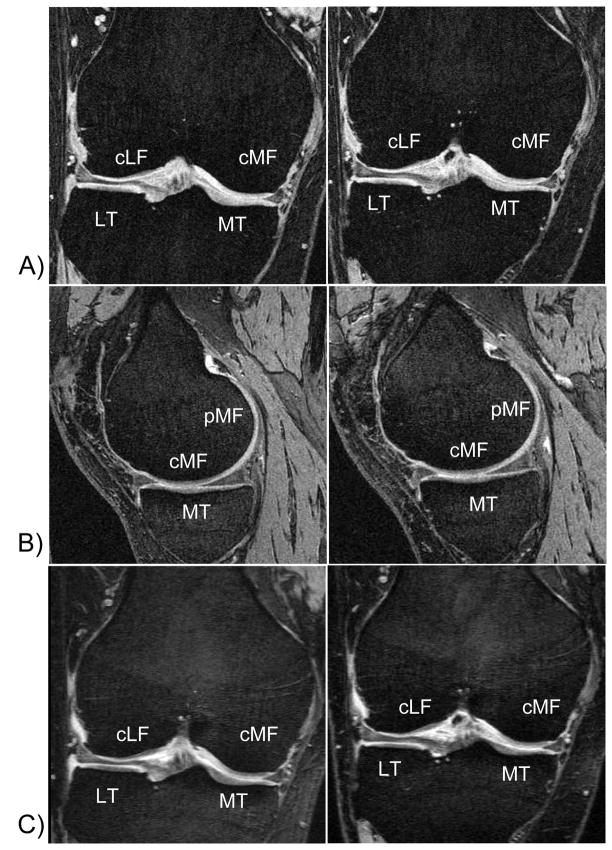
Method: Nine participants with mild to moderate clinical OA were imaged twice each at baseline, year 1 (Y1) and year 2 (Y2). Coronal 1.5mm FLASH and sagittal 0.7mm DESS sequences were acquired; 1.5mm coronal multiplanar reformats (MPR) were obtained from the DESS. Patellar, femoral and tibial cartilage plates were quantified in a paired fashion, with blinding to time point.
Results: In the weight-bearing femorotibial joint, average precision errors across plates were 1.8% for FLASH, 2.6% for DESS, and 3.0% for MPR-DESS. Volume loss at Y1 was not significant; at Y2 the average change across the femorotibial cartilage plates was -1.7% for FLASH, -2.8% for DESS, and -0.3% for MPR-DESS. Volume change in the lateral tibia (-5.5%; P<0.03), and in the medial (-2.9%; P<0.04) and lateral femorotibial compartments (-3.8%; P<0.03) were significant for DESS.
Conclusions: FLASH, DESS and MPR-DESS all displayed adequate test-retest precision. Although the comparison between protocols is limited by the small number of participants and by the relatively small longitudinal change in cartilage morphology in this pilot study, the data suggest that significant change can be detected with MRI in a small sample of OA subjects over 2 years.
Conflict of interest statement
Figures
References
-
- Peterfy CG. Imaging of the disease process. Curr Opin Rheumatol. 2002;14:590–6. - PubMed
-
- Peterfy CG. Role of MR imaging in clinical research studies. Semin Musculoskelet Radiol. 2001;5:365–78. - PubMed
-
- Gray ML, Eckstein F, Peterfy C, Dahlberg L, Kim YJ, Sorensen AG. Toward imaging biomarkers for osteoarthritis. Clin Orthop. 2004:S175–S181. - PubMed
-
- Peterfy CG, Gold G, Eckstein F, Cicuttini F, Dardzinski B, Stevens R. MRI protocols for whole-organ assessment of the knee in osteoarthritis. Osteoarthritis Cartilage. 2006;14(Suppl 1):95–111. Epub;%2006 Jun 5.:95–111. - PubMed
-
- Eckstein F, Cicuttini F, Raynauld JP, Waterton JC, Peterfy C. Magnetic resonance imaging (MRI) of articular cartilage in knee osteoarthritis (OA): morphological assessment . Osteoarthritis Cartilage. 2006;14(Suppl 1):46–75. Epub;%2006 May;%19.:46–75. - PubMed
Publication types
MeSH terms
Grants and funding
LinkOut - more resources
Full Text Sources
Medical
Research Materials
