Review
. 2007 Aug;1771(8):952-60.
doi: 10.1016/j.bbalip.2007.04.018. Epub 2007 May 22.

Modulation of PPAR activity via phosphorylation

Affiliations
Review

Modulation of PPAR activity via phosphorylation

Katherine A Burns et al. Biochim Biophys Acta. 2007 Aug.

Abstract

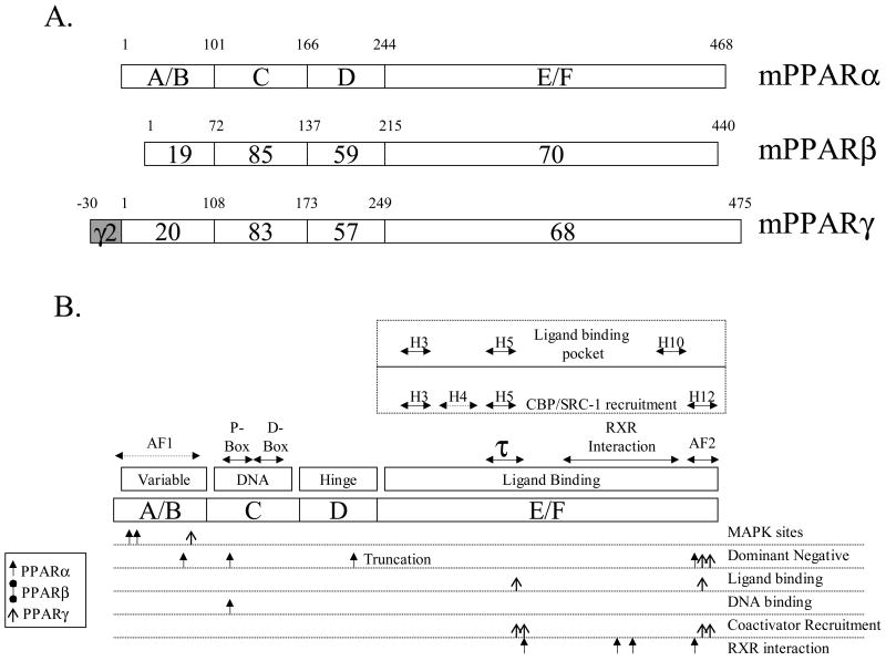
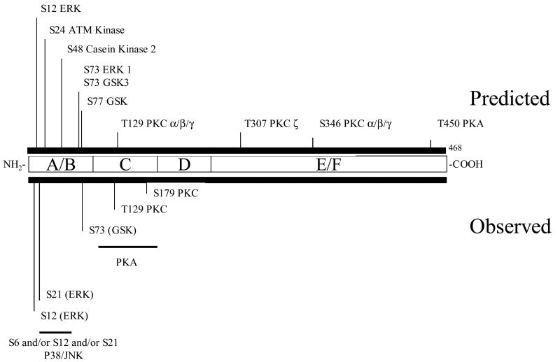
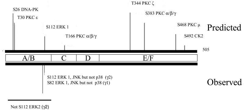
Peroxisome proliferator-activated receptors (PPARs) are members of the nuclear receptor superfamily of transcription factors that respond to specific ligands by altering gene expression in a cell-, developmental- and sex-specific manner. Three subtypes of this receptor have been discovered (PPARalpha, beta and gamma), each apparently evolving to fulfill different biological niches. PPARs control a variety of target genes involved in lipid homeostasis, diabetes and cancer. Similar to other nuclear receptors, the PPARs are phosphoproteins and their transcriptional activity is affected by cross-talk with kinases and phosphatases. Phosphorylation by the mitogen-activated protein kinases (ERK- and p38-MAPK), Protein Kinase A and C (PKA, PKC), AMP Kinase (AMPK) and glycogen synthase kinase-3 (GSK3) affect their activity in a ligand-dependent or -independent manner. The effects of phosphorylation depend on the cellular context, receptor subtype and residue metabolized which can be manifested at several steps in the PPAR activation sequence including ligand affinity, DNA binding, coactivator recruitment and proteasomal degradation. The review will summarize the known PPAR kinases that directly act on these receptors, the sites affected and the result of this modification on receptor activity.


Figures

Figure 1
Figure 1
Structure of PPARs. Panel A. Structure and functional domains of PPARα, β and γ. A/B is the hypervariable region containing the putative activation function-1 (AF-1) domain. PPARγ2 contains a 30 amino acid region that arises from differential promoter use and splicing. The C-domain is the most conserved and contains the DNA binding motif. The D-domain (hinge) is believed to allow for conformational change following ligand binding. The E/F domain contains the ligand-binding region of PPAR. Alignments and percent similarities were performed with MegAlign (DNAStar, Madison WI). Panel B. Detailed functional domains of PPARs. Above the outline for the hypothetical PPAR are the structural features of PPARs that have been deduced by sequence comparisons and crystallography. The AF-1 domain has not been fully characterized although it is known to reside in the A/B domain. The DNA binding motif contains two C4 zinc fingers, the proximal (P-box) and distal (D-box) boxes, which confer DNA binding and heterodimerization, respectively. Much has been learned about PPAR structure/function from recent crystallographic studies. PPAR E/F contains 13 alpha helices (H1-H12, H2′) and 4 short β strands [60] and helices 3, 5 and 10 forms the ligand-binding pocket. RXR interacts along several helices including H7-H10. The coactivator CBP interacts with H3-5 and H12 while SRC-1 associated with H3, H5 and H12. The τ1 domain contains a leucine-zipper-like heptad repeat [61]. The AF-2 domain is highly conserved among all PPARs and is intimately associated with ligand-induced transcriptional events. Much of the characterization of functional domains was performed using site-directed mutagenesis, as shown below the hypothetical PPAR. MAPK phosphorylation sites have been found at S12 and S21 in mouse PPARα and S122 in PPARγ2 [2]. Dominant negative (dn) PPARα results from mutations at L71, L123, and V444 [62] or in the naturally occurring truncated form of the receptor [63]. A dn PPARγ can be formed by mutating L468 and E471 of the human receptor [64]. Ligand binding mutants may arise from altering residues L319 or L469 of hPPARγ [65]. Similarly, DNA binding-devoid constructs are produced by mutating C122 [66] of PPARα. Specific interactions, such as those with SRC-1 and CBP, are targeted by mutating residues K301, V315, L468 or E471 of hPPARγ [65] while RXR association is lost by changing sites L433 [66], L370, L391 or D304 [61] of hPPARα. It is important to note that all domains work as a unified entity, with changes at the A/B terminus affecting ligand binding at the COOH, E/F domain [67] or in DNA binding [68]. Very little mutational analysis has been performed with PPARβ/γ, although the crystal structure reveals an E/F domain of this subtype to be very similar to PPARγ.
Figure 2
Figure 2. Structure of mouse PPARα and location of phosphorylation sites
Serine or threonine consensus sites are marked with their location (i.e. S12 is serine at residue 12). The key shows the font associated with each kinase's consensus site. Abbreviations used: MAPK, mitogen activated protein kinase; PKC, protein kinase C; CK2, casein kinase 2; GSK3, glycogen synthase kinase 3; PKA, protein kinase A. Consensus sites scanned using Scansite (http://scansite.mit.edu/) under moderate stringency. The sequence was scanned for the following kinase sites: Akt_Kin, ATM_Kin, Cam_Kin2, Casn_Kin1, Casn_Kin2, Cdc2_Kin, Cdk5_Kin, Clk2_Kin, DNA_PK, Erk1_Kin, GSK3_Kin, p38_Kin, PKA_Kin, PKC_common, PKC_delta, PKC_epsilon, PKC_mu, PKC_zeta.
Figure 3
Figure 3. Structure of mouse PPARβ/δ and location of phosphorylation sites
See Figure 2 for details.
Figure 4
Figure 4. Structure of mouse PPARγ2 and location of phosphorylation sites
See Figure 2 for details.

References

    1. N.R.N. Committee. A unified nomenclature system for the nuclear receptor superfamily. Cell. 1999;97:161–163. - PubMed
    1. Escher P, Wahli W. Peroxisome proliferator-activated receptors: insight into multiple cellular functions. Mutat Res. 2000;448:121–138. - PubMed
    1. Braissant O, Foufelle F, Scotto C, Dauca M, Wahli W. Differential expression of peroxisome proliferator-activated receptors (PPARs): tissue distribution of PPAR-alpha, - beta, and -gamma in the adult rat. Endocrinology. 1996;137:354–366. - PubMed
    1. Fajas L, Auboeuf D, Raspe E, Schoonjans K, Lefebvre AM, Saladin R, Najib J, Laville M, Fruchart JC, Deeb S, Vidal-Puig A, Flier J, Briggs MR, Staels B, Vidal H, Auwerx J. The organization, promoter analysis, and expression of the human PPARgamma gene. J Biol Chem. 1997;272:18779–18789. - PubMed
    1. Issemann I, Green S. Activation of a member of the steroid hormone receptor superfamily by peroxisome proliferators. Nature. 1990;347:645–650. - PubMed

Publication types

MeSH terms