Review
. 2008 Feb 4:1192:29-36.
doi: 10.1016/j.brainres.2007.05.021. Epub 2007 May 23.

Nuclear migration during retinal development

Affiliations
Review

Nuclear migration during retinal development

Lisa M Baye et al. Brain Res. .

Abstract

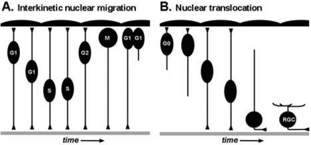
In this review we focus on the mechanisms, regulation, and cellular consequences of nuclear migration in the developing retina. In the nervous system, nuclear migration is prominent during both proliferative and post-mitotic phases of development. Interkinetic nuclear migration is the process where the nucleus oscillates from the apical to basal surfaces in proliferative neuroepithelia. Proliferative nuclear movement occurs in step with the cell cycle, with M-phase being confined to the apical surface and G1-, S-, and G2-phases occurring at more basal locations. Later, following cell cycle exit, some neuron precursors migrate by nuclear translocation. In this mode of cellular migration, nuclear movement is the driving force for motility. Following discussion of the key components and important regulators for each of these processes, we present an emerging model where interkinetic nuclear migration functions to distinguish cell fates among retinal neuroepithelia.


Figures

Figure 1
Figure 1. Types of nuclear migration during retinal development
(A) Model of interkinetic nuclear migration. As cells progress through the cell cycle, nuclei of neuroepithelial cells move in an apical-to-basal and then basal-to-apical fashion. M-phase is restricted to the apical surface near the RPE. Note the maintenance of both apical and basal processes throughout the cell cycle. With cytokinesis, typically one daughter cell inherits the basal process and the sibling cell initiates a new basal process. (B) Model of nuclear translocation. Following cell cycle exit, progenitors that migrate via nuclear translocation maintain their apical and basal processes. These processes retract as the soma reaches the appropriate laminar position. Apical RPE is up and basal basement membrane is down.
Figure 2
Figure 2. Motor and adaptor proteins for nuclear migration
(A) Model of interkinetic nuclear migration and coordination with the cell cycle. In some contexts, directed nuclear migration is a balance between the relative activity of plus-end (dark blue) and minus-end (orange) oriented microtubule motor proteins. (B) Model showing proteins known to function during nuclear anchoring and nuclear migration in various contexts. SUN-domain proteins (red); KASH-domain proteins (green); Dynein-dynactin (orange) with associated Arp proteins (grey). Nucleus (blue) is shown with the nuclear lamina (grey). ONM, outer nuclear membrane; INM, inner nuclear membrane; NPC, Nucleopore complex. See text for details regarding each cartoon.
Figure 3
Figure 3. Interkinetic nuclear migration and cell fate diversification
(A) Model showing localized extrinsic (right side) and intrinsic signals (left). Evidence exists for ATP released by the RPE, Notch activating components at the apical side of the neuroepithelium, and various soluble factors released from post-mitotic retinal ganglion cells. Within retinal neuroepithelial cells, distinct signaling complexes reside at the apical and basal poles (Apical junctional complex, red; Focal adhesion complex, blue). (B) Heterogeneity in the position of G1-phase (green nuclei), S-phase (dark blue), and G2-phase (light blue) during interkinetic nuclear migration. M-phase is always restricted to the apical surface (yellow nuclei). In this example, cells in G1-phase are competent to respond to localized intrinsic and/or extrinsic signals that promote a red-cell fate. Because of differential nuclear positions, subsets of neuroepithelia (red cells) diverge in fate from others (grey cells). Note in this example, only G1 cells with more basal nuclei undergo the red cell fate commitment. Apical RPE is up and basal basement membrane is down.

References

    1. Apel ED, Lewis RM, Grady RM, Sanes JR. Syne-1, a dystrophin- and Klarsicht-related protein associated with synaptic nuclei at the neuromuscular junction. J Biol Chem. 2000;275:31986–95. - PubMed
    1. Baye LM, Link BA. Examining interkinetic nuclear migration during retinal development. FASEB JOURNAL. 2005;19:A1367. abstract.
    1. Baye LM, Link BA. The disarrayed mutation results in cell cycle and neurogenesis defects during retinal development in zebrafish. BMC Dev Biol. 2007;7:28. - PMC - PubMed
    1. Beggs HE, Schahin-Reed D, Zang K, Goebbels S, Nave KA, Gorski J, Jones KR, Sretavan D, Reichardt LF. FAK deficiency in cells contributing to the basal lamina results in cortical abnormalities resembling congenital muscular dystrophies. Neuron. 2003;40:501–14. - PMC - PubMed
    1. Bhide PG. Cell cycle kinetics in the embryonic mouse corpus striatum. J Comp Neurol. 1996;374:506–22. - PubMed

Publication types