. 2007;8(6):R107.
doi: 10.1186/gb-2007-8-6-r107.

Large-scale identification of human genes implicated in epidermal barrier function

Affiliations

Large-scale identification of human genes implicated in epidermal barrier function

Eve Toulza et al. Genome Biol. 2007.

Abstract

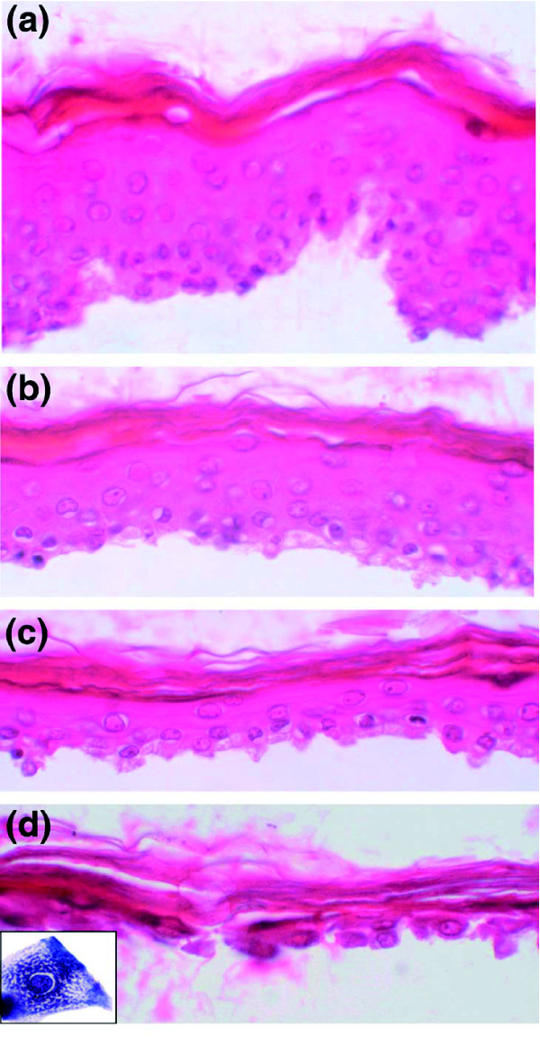
Background: During epidermal differentiation, keratinocytes progressing through the suprabasal layers undergo complex and tightly regulated biochemical modifications leading to cornification and desquamation. The last living cells, the granular keratinocytes (GKs), produce almost all of the proteins and lipids required for the protective barrier function before their programmed cell death gives rise to corneocytes. We present here the first analysis of the transcriptome of human GKs, purified from healthy epidermis by an original approach.

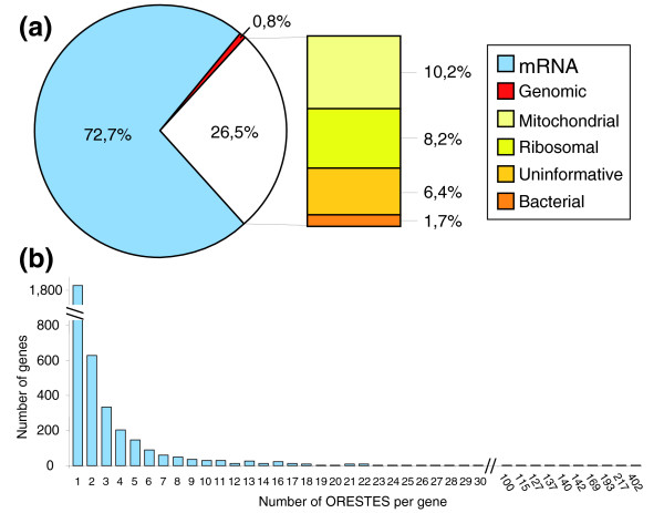
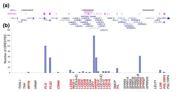
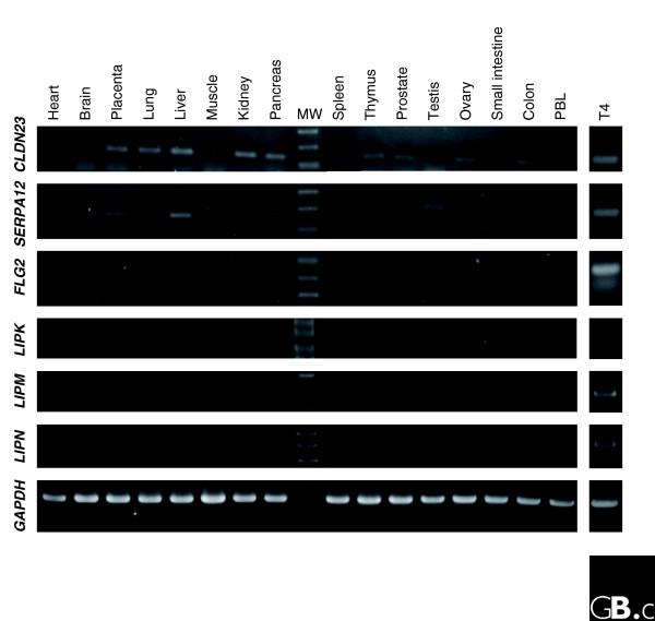
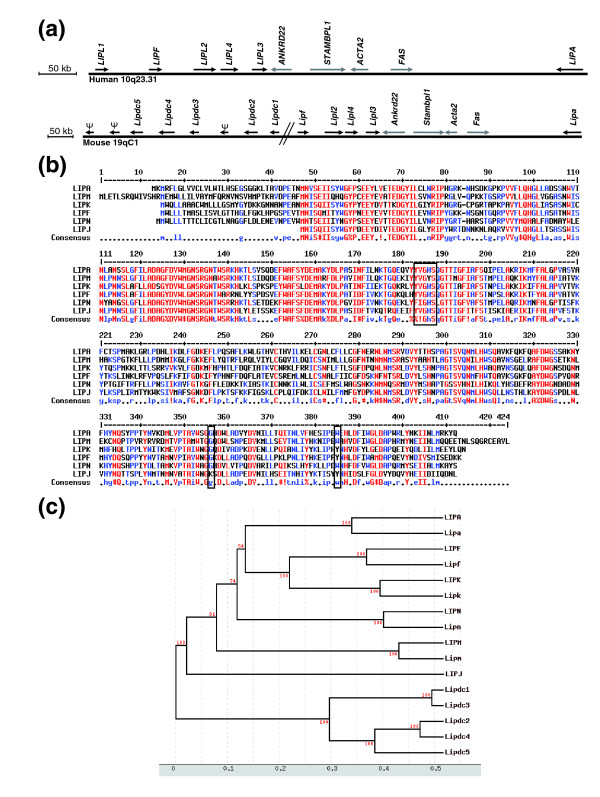
Results: Using the ORESTES method, 22,585 expressed sequence tags (ESTs) were produced that matched 3,387 genes. Despite normalization provided by this method (mean 4.6 ORESTES per gene), some highly transcribed genes, including that encoding dermokine, were overrepresented. About 330 expressed genes displayed less than 100 ESTs in UniGene clusters and are most likely to be specific for GKs and potentially involved in barrier function. This hypothesis was tested by comparing the relative expression of 73 genes in the basal and granular layers of epidermis by quantitative RT-PCR. Among these, 33 were identified as new, highly specific markers of GKs, including those encoding a protease, protease inhibitors and proteins involved in lipid metabolism and transport. We identified filaggrin 2 (also called ifapsoriasin), a poorly characterized member of the epidermal differentiation complex, as well as three new lipase genes clustered with paralogous genes on chromosome 10q23.31. A new gene of unknown function, C1orf81, is specifically disrupted in the human genome by a frameshift mutation.

Conclusion: These data increase the present knowledge of genes responsible for the formation of the skin barrier and suggest new candidates for genodermatoses of unknown origin.


Figures

Figure 1
Figure 1
Histological analysis of epidermis samples. (a) Hematoxylin-eosin stained sections of entire epidermis after thermolysin incubation and removal of the dermis. (b,c,d) Epidermis fragments remaining after the first, second, and third trypsin incubation, respectively. Fragments shown in (d) are mainly composed of GKs attached to the cornified layer and constitute the T4 fraction. Inset: higher magnification showing the characteristic cytological aspect of a GK with cytoplasmic keratohyalin granules.
Figure 2
Figure 2
Analysis of the ORESTES dataset from GKs. (a) Pie graph of the 22,585 sequences obtained from the T4 fraction enriched in GKs. The treatment of the mRNA samples with DNAse resulted in minimal contamination with genomic sequences. Despite two rounds of polyA+ mRNA purification, rRNA sequences still represent approximately 8% of the dataset. (b) Histogram showing the number of ORESTES at each level of redundancy. The vast majority of genes are represented by less than five ORESTES, illustrating the normalization capability of that method. However, a small number of genes are represented by a large number of ORESTES (up to 402).
Figure 3
Figure 3
Genes of the EDC present in the ORESTES dataset. (a) Screen copy of a UCSC Genome Browser window (chr1:150,300,000-151,590,000; hg17, May 2004) showing the RefSeq genes from the EDC, and the ORESTE custom track. (b) Number of ORESTES for each gene of the locus. The genes for which at least one ORESTE was sequenced are in red bold characters.
Figure 4
Figure 4
Expression profile of newly identified genes. PCR experiments were performed with a commercial panel of cDNAs from 16 human tissues (PBL, peripheral blood leukocytes) and with cDNAs prepared from the T4 fraction enriched in GKs. For each gene, PCR primers were chosen to amplify a cDNA fragment encompassing at least two exons. Note the highly specific expression pattern of FLG2, LIPK, M, N, and, to a lesser extent, SERPINA12 genes. The apparent size variation of the CLDN23 fragment results from an artifactual gel distortion. Expression of GAPDH, assessed with the primers provided by the manufacturer, was used as a control.
Figure 5
Figure 5
Analysis of new human lipase genes. (a) Schematic representation of the lipase gene cluster on chromosome 10q23.31. Six lipase genes, including the newly described LIPL1 (LIPJ), LIPL2 (LIPK), LIPL3 (LIPM) and LIPL4 (LIPN), form a cluster also containing four unrelated genes. (b) Alignment of the protein sequences of the six human lipases from the chromosome 10q23.31 cluster. The amino acids of the catalytic triad are boxed. The alignment was generated with Multalin software [71]. (c) Hierarchical clustering of human and mouse abhydro-lipase gene family members. The human LIPA, LIPF, LIPK, LIPM and LIPN, but not LIPJ, proteins have clear mouse orthologues (lower-case gene names). The six hypothetical mouse genes found in place of the LIPJ gene (Lipdc1-5) form a separate branch of the phylogenetic tree. This tree was generated with the Tree Top software [72]. Bootstrap values are indicated in red.
Figure 6
Figure 6
C1orf81 mRNA expression and conservation of the eighth exon among mammals. (a) Expression pattern of the C1orf81 gene. PCR was performed with a commercial panel of cDNAs from 16 human tissues (PBL, peripheral blood leukocytes) and with cDNA prepared from epidermis. The amplified fragment (120 nt) encompasses exons 13-14. (b) Sequence alignment of the eighth exon of the C1orf81 gene from 11 mammals. The sequences were retrieved from the multiz17way table of the UCSC Genome Browser [73], and from a BLAST search of the cat genome. The consensus splicing signals are boxed. The black arrow indicates the single nucleotide insertion in the human gene. The alignment was created with Multalin software [71]. Asterisks indicate the positions conserved in all the sequences. The colors correspond to various levels of consensus, with red for high consensus and grey for low consensus.

References

    1. Watt FM, Celso CL, Silva-Vargas V. Epidermal stem cells: an update. Curr Opin Genet Dev. 2006;16:518–524. doi: 10.1016/j.gde.2006.08.006. - DOI - PubMed
    1. Park GT, Lim SE, Jang SI, Morasso MI. Suprabasin, a novel epidermal differentiation marker and potential cornified envelope precursor. J Biol Chem. 2002;277:45195–45202. doi: 10.1074/jbc.M205380200. - DOI - PMC - PubMed
    1. Oomizu S, Sahuc F, Asahina K, Inamatsu M, Matsuzaki T, Sasaki M, Obara M, Yoshizato K. Kdap, a novel gene associated with the stratification of the epithelium. Gene. 2000;256:19–27. doi: 10.1016/S0378-1119(00)00357-7. - DOI - PubMed
    1. Makino T, Takaishi M, Morohashi M, Huh NH. Hornerin, a novel profilaggrin-like protein and differentiation-specific marker isolated from mouse skin. J Biol Chem. 2001;276:47445–47452. doi: 10.1074/jbc.M107512200. - DOI - PubMed
    1. Lee WH, Jang S, Lee JS, Lee Y, Seo EY, You KH, Lee SC, Nam KI, Kim JM, Kee SH, et al. Molecular cloning and expression of human keratinocyte proline-rich protein (hKPRP), an epidermal marker isolated from calcium-induced differentiating keratinocytes. J Invest Dermatol. 2005;125:995–1000. doi: 10.1111/j.0022-202X.2005.23829.x. - DOI - PubMed

Publication types

LinkOut - more resources