. 2007 Jul 9;204(7):1509-17.
doi: 10.1084/jem.20061675. Epub 2007 Jun 11.

Interleukin 25 promotes the initiation of proallergic type 2 responses

Affiliations

Interleukin 25 promotes the initiation of proallergic type 2 responses

Pornpimon Angkasekwinai et al. J Exp Med. .

Abstract

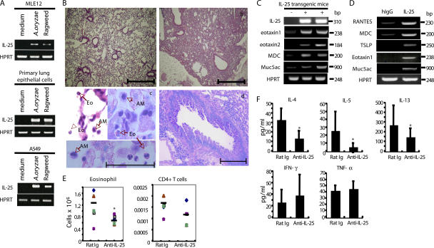
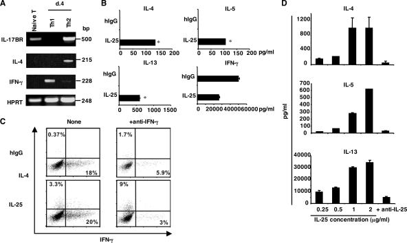
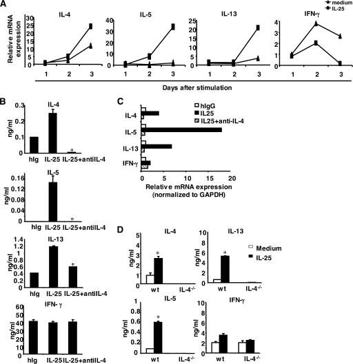
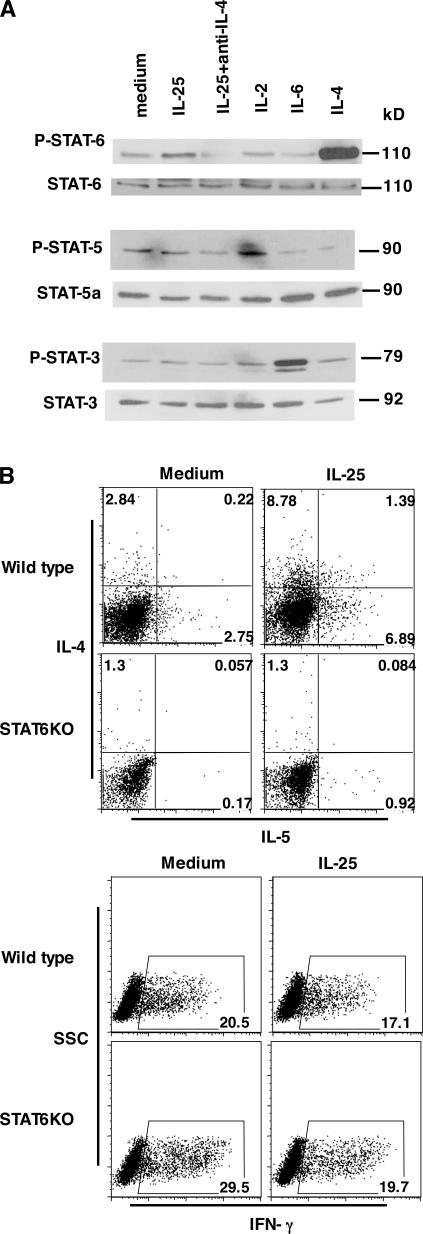
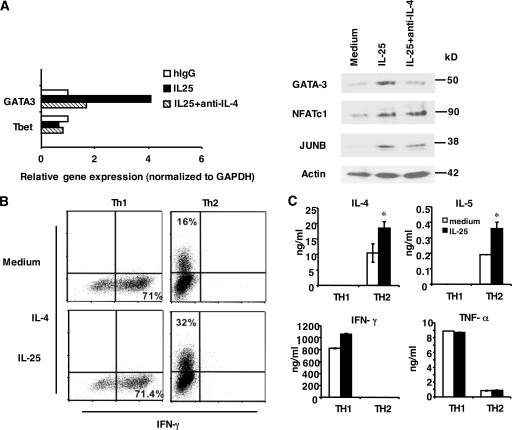
The molecular mechanisms underlying the initiation of innate and adaptive proallergic type 2 responses are not understood. Interleukin (IL) 25, a member of the IL-17 cytokine family, was recently reported (Owyang, A.M., C. Zaph, E.H. Wilson, K.J. Guild, T. McClanahan, H.R. Miller, D.J. Cua, M. Goldschmidt, C.A. Hunter, R.A. Kastelein, and D. Artis. 2006. J. Exp. Med. 203:843-849; Fallon, P.G., S.J. Ballantyne, N.E. Mangan, J.L. Barlow, A. Dasvarma, D.R. Hewett, A. McIlgorm, H.E. Jolin, and A.N. McKenzie. 2006. J. Exp. Med. 203:1105-1116) to be important in Th2 cell-mediated immunity to parasitic infection. However, the cellular source and targets of IL-25 are not well understood. We show that mouse IL-25 is expressed by lung epithelial cells as a result of innate immune responses to allergens. Transgenic overexpression of IL-25 by these cells leads to mucus production and airway infiltration of macrophages and eosinophils, whereas blockade of IL-25 conversely reduces the airway inflammation and Th2 cytokine production in an allergen-induced asthma model. In addition, IL-25, with a receptor more highly expressed in Th2 than other effector T cells, promotes Th2 cell differentiation in an IL-4- and signal transducer and activator of transcription 6-dependent manner. During early T cell activation, IL-25 potentiates expression of the nuclear factor of activated T cells c1 and JunB transcription factors, which possibly results in increased levels of initial IL-4 production, up-regulation of GATA-3 expression, and enhanced Th2 cell differentiation. Thus, IL-25 is a critical factor regulating the initiation of innate and adaptive proallergic responses.


Figures

Figure 1.
Figure 1.
IL-25 is expressed by lung epithelial cells and regulates allergic responses. (A) MLE12 cells, A549 cells, and primary type II alveolar epithelial cells stimulated with the indicated stimuli were analyzed by RT-PCR for IL-25 mRNA expression after normalization with HPRT expression. (B) Histological analysis of lung tissues from 3-mo-old IL-25 transgenic mice (b) stained with H&E was compared with that from control littermate mice (a). Bars, 500 μm. Macrophages (AM) and eosinophils (Eo) infiltrated in the airway were demonstrated by H&E and Giemsa staining (c), and mucus hyperplasia stained with PAS in 5-mo-old IL-25 transgenic-positive mice are shown (d). Bars: (c) 50 μm; (d) 200 μm. (C) RT-PCR analysis of gene expression in 3-mo-old IL-25 transgenic mice was compared with littermate controls. (D) MLE12 cells treated for 6 h with IL-25 were subject to RT-PCR analysis in comparison with the nontreated cells. (E and F) C57BL/6 mice (4–5 mice in each group) were intranasally challenged with A. oryzae allergen and OVA every other day for a total of six challenges. An anti–IL-25 mAb or control rat Ig was intraperitoneally administered at the time of each challenge. 24 h after the final challenging, mice were killed, and BAL cells were collected and analyzed for the absolute numbers of eosinophils and CD4+ T cells by total cell counting, differential cell counts, and flow cytometry analysis with mAbs to CCR3 (for eosinophils) and CD4 (BD Biosciences). BAL fluid was examined for cytokine expression by ELISA. Horizontal bars in E represent the means. Data are presented as mean values + SD and are representative of two independent experiments. HPRT, hypoxanthine-guanine phosphoribosyltransferase. *, P < 0.05.
Figure 2.
Figure 2.
IL-25 promotes Th2 cell differentiation in vitro. (A) mRNA expression of IL-17RB on freshly isolated naive T cells and in vitro–generated Th1 and Th2 cells was analyzed by RT-PCR. (B) Naive T cells were stimulated with anti-CD3, anti-CD28, and IL-2 in the presence of human IgG or recombinant IL-25. After 4 d, cells were restimulated with plate-bound anti-CD3 for 24 h, and cytokine production was measured by ELISA. *, P < 0.005. (C) Naive T cells were activated as described with or without anti–IFN-γ in the presence of human IgG or recombinant IL-25. After 5 d, cells were restimulated with PMA/ionomycin, and the expression of IL-4 and IFN-γ was analyzed. Numbers within the quadrants indicate the percentage of cells stained positive for each respective cytokine. (D) Naive T cells were purified and stimulated with anti-CD3, anti-CD28, anti–IFN-γ, and IL-2 in the presence of different concentrations of IL-25 for 6 d. 50 μg/ml anti–IL-25 mAb was added to the culture containing IL-25 at 2 μg/ml. The amounts of cytokines were measured by ELISA. Data are presented as mean values + SD and are representative of at least two independent experiments. HPRT, hypoxanthine-guanine phosphoribosyltransferase.
Figure 3.
Figure 3.
IL-25 promotes Th2 cell differentiation in an IL-4-dependent manner. (A) Naive T cells were purified and stimulated with anti-CD3, anti-CD28, and IL-2 with or without IL-25. Cells were collected at the indicated time points for real-time PCR analysis. (B and C) Naive T cells were stimulated with anti-CD3, anti-CD28, and IL-2 with or without IL-25 or with the addition of anti–IL-4 mAb. After 4 d of culture, cells were washed and restimulated with anti-CD3 for 4 h to analyze the cytokine expression by real-time RT-PCR or for 24 h to measure the level of cytokine expression by ELISA. *, P < 0.05. (D) Naive T cells from IL-4–deficient mice were stimulated as described with or without IL-25 and restimulated to measure the level of cytokine expression by ELISA. Data are presented as mean values + SD and are representative of two experiments. *, P < 0.05.
Figure 4.
Figure 4.
IL-25 promotes Th2 cell differentiation in a STAT6-dependent manner. (A) Naive T cells were purified and stimulated with anti-CD3 and anti-CD28 with or without IL-25. IL-4, IL-2, and IL-6 were included in the treatment as positive controls for STAT6, STAT5, and STAT3 phosphorylation, respectively. Cells were collected at 48 h for Western blot analysis. (B) Purified naive T cells from STAT6-deficient and wild-type mice (BALB/c) were stimulated with anti-CD3, anti-CD28, and IL-2 with or without IL-25. After 5 d of culture, cells were restimulated with PMA/ionomycin to assess the levels of IL-4 and IL-5 and IFN-γ expression. Numbers in the quadrants/gates represent the percentage of cells stained positive for each respective cytokine. SSC, side scatter.
Figure 5.
Figure 5.
The function of IL-25 in early Th cell activation and in effector Th2 cells. (A) Naive T cells were stimulated with anti-CD3 and anti-CD28 with or without IL-25 or with the addition of anti–IL-4 antibody. Cells were collected at 72 h for real-time PCR or 48 h for Western blot analysis. (B) Purified naive T cells from transgenic OT-II mice were differentiated into Th1 and Th2 cells. After 5 d of culture, cells were washed and restimulated with anti-CD3 with or without IL-25 for 3 d to assess the levels of IL-4 and IFN-γ expression by intracellular cytokine staining. The percentages in the figure represent cells stained positive for each cytokine indicated. (C) Purified naive T cells from C57BL/6 mice were stimulated with anti-CD3 and anti-CD28 with the same treatment as in B. After 5 d of culture, Th1 and Th2 cells were washed and restimulated with anti-CD3 with or without IL-25 for 2 d to assess the levels of IL-4, IL-5, IFN-γ, and TNF-α expression by ELISA. Data are presented as mean values + SD. *, P < 0.05.

References

    1. Murphy, K.M. 1998. T lymphocyte differentiation in the periphery. Curr. Opin. Immunol. 10:226–232. - PubMed
    1. Dong, C., and R.A. Flavell. 2000. Cell fate decision: T-helper 1 and 2 subsets in immune responses. Arthritis Res. 2:179–188. - PMC - PubMed
    1. Dong, C. 2006. Diversification of T-helper-cell lineages: finding the family root of IL-17-producing cells. Nat. Rev. Immunol. 6:329–334. - PubMed
    1. Dong, C., and R.A. Flavell. 2000. Control of T helper cell differentiation—in search of master genes. Sci. STKE. 2000:PE1. - PubMed
    1. Hurst, S.D., T. Muchamuel, D.M. Gorman, J.M. Gilbert, T. Clifford, S. Kwan, S. Menon, B. Seymour, C. Jackson, T.T. Kung, et al. 2002. New IL-17 family members promote Th1 or Th2 responses in the lung: in vivo function of the novel cytokine IL-25. J. Immunol. 169:443–453. - PubMed