

Caenorhabditis elegans SMG-2 selectively marks mRNAs containing premature translation termination codons
- PMID: 17562857
- PMCID: PMC1952128
- DOI: 10.1128/MCB.00410-07
Caenorhabditis elegans SMG-2 selectively marks mRNAs containing premature translation termination codons
Abstract
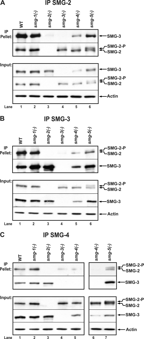
Eukaryotic mRNAs containing premature translation termination codons (PTCs) are rapidly degraded by a process termed "nonsense-mediated mRNA decay" (NMD). We examined protein-protein and protein-RNA interactions among Caenorhabditis elegans proteins required for NMD. SMG-2, SMG-3, and SMG-4 are orthologs of yeast (Saccharomyces cerevisiae) and mammalian Upf1, Upf2, and Upf3, respectively. A combination of immunoprecipitation and yeast two-hybrid experiments indicated that SMG-2 interacts with SMG-3, SMG-3 interacts with SMG-4, and SMG-2 interacts indirectly with SMG-4 via shared interactions with SMG-3. Such interactions are similar to those observed in yeast and mammalian cells. SMG-2-SMG-3-SMG-4 interactions require neither SMG-2 phosphorylation, which is abolished in smg-1 mutants, nor SMG-2 dephosphorylation, which is reduced or eliminated in smg-5 mutants. SMG-2 preferentially associates with PTC-containing mRNAs. We monitored the association of SMG-2, SMG-3, and SMG-4 with mRNAs of five endogenous genes whose mRNAs are alternatively spliced to either contain or not contain PTCs. SMG-2 associates with both PTC-free and PTC-containing mRNPs, but it strongly and preferentially associates with ("marks") those containing PTCs. SMG-2 marking of PTC-mRNPs is enhanced by SMG-3 and SMG-4, but SMG-3 and SMG-4 are not detectably associated with the same mRNPs. Neither SMG-2 phosphorylation nor dephosphorylation is required for selective association of SMG-2 with PTC-containing mRNPs, indicating that SMG-2 is phosphorylated only after premature terminations have been discriminated from normal terminations. We discuss these observations with regard to the functions of SMG-2 and its phosphorylation during NMD.
Figures


Similar articles
-
SMG-2 is a phosphorylated protein required for mRNA surveillance in Caenorhabditis elegans and related to Upf1p of yeast.Mol Cell Biol. 1999 Sep;19(9):5943-51. doi: 10.1128/MCB.19.9.5943. Mol Cell Biol. 1999. PMID: 10454541 Free PMC article.
-
SMG-1 is a phosphatidylinositol kinase-related protein kinase required for nonsense-mediated mRNA Decay in Caenorhabditis elegans.Mol Cell Biol. 2004 Sep;24(17):7483-90. doi: 10.1128/MCB.24.17.7483-7490.2004. Mol Cell Biol. 2004. PMID: 15314158 Free PMC article.
-
The Substrates of Nonsense-Mediated mRNA Decay in Caenorhabditis elegans.G3 (Bethesda). 2018 Jan 4;8(1):195-205. doi: 10.1534/g3.117.300254. G3 (Bethesda). 2018. PMID: 29122854 Free PMC article.
-
Role of SMG-1-mediated Upf1 phosphorylation in mammalian nonsense-mediated mRNA decay.Genes Cells. 2013 Mar;18(3):161-75. doi: 10.1111/gtc.12033. Epub 2013 Jan 28. Genes Cells. 2013. PMID: 23356578 Review.
-
The role of SMG-1 in nonsense-mediated mRNA decay.Biochim Biophys Acta. 2005 Dec 30;1754(1-2):305-15. doi: 10.1016/j.bbapap.2005.10.002. Epub 2005 Oct 25. Biochim Biophys Acta. 2005. PMID: 16289965 Review.
Cited by
-
Target Discrimination in Nonsense-Mediated mRNA Decay Requires Upf1 ATPase Activity.Mol Cell. 2015 Aug 6;59(3):413-25. doi: 10.1016/j.molcel.2015.06.036. Mol Cell. 2015. PMID: 26253027 Free PMC article.
-
Is there a classical nonsense-mediated decay pathway in trypanosomes?PLoS One. 2011;6(9):e25112. doi: 10.1371/journal.pone.0025112. Epub 2011 Sep 21. PLoS One. 2011. PMID: 21957477 Free PMC article.
-
Nonsense-Mediated mRNA Decay Begins Where Translation Ends.Cold Spring Harb Perspect Biol. 2019 Feb 1;11(2):a032862. doi: 10.1101/cshperspect.a032862. Cold Spring Harb Perspect Biol. 2019. PMID: 29891560 Free PMC article. Review.
-
The role of nucleotide composition in premature termination codon recognition.BMC Bioinformatics. 2016 Dec 7;17(1):519. doi: 10.1186/s12859-016-1384-z. BMC Bioinformatics. 2016. PMID: 27927164 Free PMC article.
-
A gripping tale of ribosomal frameshifting: extragenic suppressors of frameshift mutations spotlight P-site realignment.Microbiol Mol Biol Rev. 2009 Mar;73(1):178-210. doi: 10.1128/MMBR.00010-08. Microbiol Mol Biol Rev. 2009. PMID: 19258537 Free PMC article. Review.
References
-
- Alonso, C. R. 2005. Nonsense-mediated RNA decay: a molecular system micromanaging individual gene activities and suppressing genomic noise. Bioessays 27:463-466. - PubMed
-
- Amrani, N., R. Ganesan, S. Kervestin, D. A. Mangus, S. Ghosh, and A. Jacobson. 2004. A faux 3′-UTR promotes aberrant termination and triggers nonsense-mediated mRNA decay. Nature 432:112-118. - PubMed
-
- Arciga-Reyes, L., L. Wootton, M. Kieffer, and B. Davies. 2006. UPF1 is required for nonsense-mediated mRNA decay (NMD) and RNAi in Arabidopsis. Plant J. 47:480-489. - PubMed
-
- Aronoff, R., R. Baran, and J. Hodgkin. 2001. Molecular identification of smg-4, required for mRNA surveillance in C. elegans. Gene 268:153-164. - PubMed
Publication types
MeSH terms
Substances
Grants and funding
LinkOut - more resources
Full Text Sources
Molecular Biology Databases