. 2007 Aug;27(16):5765-75.
doi: 10.1128/MCB.01848-06. Epub 2007 Jun 11.

Nuclear factor 1 and T-cell factor/LEF recognition elements regulate Pitx2 transcription in pituitary development

Affiliations

Nuclear factor 1 and T-cell factor/LEF recognition elements regulate Pitx2 transcription in pituitary development

Di Ai et al. Mol Cell Biol. 2007 Aug.

Abstract

Pitx2, a paired-related homeobox gene that is mutated in Rieger syndrome I, is the earliest known marker of oral ectoderm. Pitx2 was previously shown to be required for tooth, palate, and pituitary development in mice; however, the mechanisms regulating Pitx2 transcription in the oral ectoderm are poorly understood. Here we used an in vivo transgenic approach to investigate the mechanisms regulating Pitx2 transcription. We identified a 7-kb fragment that directs LacZ expression in oral ectoderm and in many of its derivatives. Deletion analysis of transgenic embryos reduced this fragment to a 520-bp region that directed LacZ activity to Rathke's pouch. A comparison of the mouse and human sequences revealed a conserved nuclear factor 1 (NF-1) recognition element near a consensus T-cell factor (TCF)/LEF binding site. The mutation of either site individually abolished LacZ activity in transgenic embryos, identifying Pitx2 as a direct target of Wnt signaling in pituitary development. These findings uncover a requirement for NF-1 and TCF factors in Pitx2 transcriptional regulation in the pituitary and provide insight into the mechanisms controlling region-specific transcription in the oral ectoderm and its derivatives.


Figures

FIG. 1.
FIG. 1.
Identification of a Pitx2c oral ectoderm and RP enhancer. Shown at the top of panel A is a representation of the genomic fragment that covers the Pitx2c promoter region (P2); exons 4, 5, and 6; and flanking regions. Construct names are indicated on the left, and the corresponding expression patterns are summarized on the right. The “# with expression” column shows the number of transgenic lines with positive RP and oral ectoderm staining per total number of transgenic lines. Asterisks indicate stable lines that were analyzed in multiple stages. Founder embryos were analyzed in other lines. In constructs tg1k to tg3k-Del:5812-9918, a promoterless lacZ cassette was inserted into the 5′ untranscribed region of exon4, so that it was controlled by Pitx2c regulatory elements. Construct tg1k spans Pitx2c with about 6 kb of upstream flanking sequence and 4 kb of downstream flanking sequence. Construct tg3k was generated by adding a 7-kb genomic fragment (blue line) downstream to tg1k. Construct tg2k was generated by deleting a 3-kb fragment upstream of P2 from tg3k. Construct tg3k-Del:5812-9918 was generated by deleting a 4-kb fragment in the middle of a 7-kb fragment in tg3k. Numbers (0k through 12k) show the relative location of the deleted fragment downstream to exon 6. Expression patterns from tg1k, -2k, and -3k indicated that the 7-kb fragment is essential for Pitx2c pituitary expression and the pattern from tg3k-Del:5812-9918 indicates that the deleted 4-kb fragment is dispensable for Pitx2c pituitary expression. (B) Ventral-caudal view of Pitx2c in situ hybridization of the head of a wild-type embryo (11.5 dpc). The mandible was cut off and is shown in panel C. Arrows indicate oral ectoderm and RP hybridization signals. (D to K) Whole-mount X-gal staining and sagittal sections of different stages of tg3k transgenic embryos. (D and E) X-gal staining of tg3k embryos at 11.5 dpc. The mandible was cut off and is shown in panel E. Arrows indicate LacZ staining in RP and oral ectoderm with LacZ absence in the ectoderm of the frontonasal process and most distal mandible (dotted line). (F) Sagittal section of an embryo (11.5 dpc) showing LacZ in RP (arrow). (G to H) Whole-mount X-gal staining of oral cavities of tg3k embryos at 14.5 dpc (G) and 13.5 dpc (H). Sagittal sections showed that LacZ was expressed in molar tooth (I), pituitary (J), and tongue epithelium (K) in 13.5-dpc embryos. Abbreviations: fn, frontal nasal process; md, mandible; di, diencephalon; ru, rugae; ps, palatal shelf; t, tongue; p, pituitary; mo, molor; mc, meckel cartilage. −, absence of oral ectoderm expression; +, presence of oral ectoderm expression.
FIG. 2.
FIG. 2.
Localization of the Pitx2c RP enhancer. (A) Construct names are indicated on the left, and the corresponding expression patterns are summarized on the right. The “# with expression” column shows the number of transgenic lines with positive RP and oral ectoderm staining per total number of transgenic lines. Constructs tg4k, -5k, and -6k were generated by fusing fragments from the 7-kb fragment to the Hsp68 LacZ reporter. X-gal expression of tg4k, -5k, and -6k indicated that the first 1.8-kb fragment of 7 kb was sufficient to initiate Pitx2c expression in RP. (B to H) Whole-mount X-gal staining of heads (11.5 dpc) of mice from transgenic lines, with mandibles removed. Constructs are labeled. Arrows denote RP, and asterisks denote no LacZ staining in RP. −, absence of RP expression; +, presence of RP expression.
FIG. 3.
FIG. 3.
Fine mapping of Pitx2c RP enhancer. (A) A 1.8-kb Pitx2 downstream fragment was fused to Hsp68 LacZ to generate tg5k. Different regions of a 1.8-kb fragment were fused to Hsp68 LacZ to generate constructs tg5k-1 through tg5k-6. In the nucleotides column, the relative location in or deletion from tg5k of each construct is shown. (B to E) Whole-mount X-gal staining of heads (11.5 dpc) of mice from transgenic lines, with mandibles removed and constructs labeled. >, sense orientation; <, antisense orientation. Arrows denote RP, and asterisks denote no LacZ staining in RP. −, absence of RP expression; +, presence of RP expression.
FIG. 4.
FIG. 4.
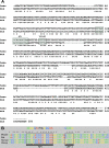
Multispecies alignment of NF-1 and LEF-1 binding sites in Pitx2c pituitary enhancer. (A) One conserved NF-1 and one TCF/LEF binding site were identified in tg5k-1 between mouse (mus) and human (homo). (B) NF-1 and TCF/LEF binding site alignments among more species. Asterisks indicate conserved nucleotides.
FIG. 5.
FIG. 5.
NF-1X and LEF-1 bind to the Pitx2c RP enhancer in vitro. (A) Schematic representation of the 525-bp minimal Pitx2c pituitary enhancer region (tg5k-1). γ-32P-labeled oligonucleotides for the NF-1 binding site and LEF-1 binding site of the Pitx2c pituitary regulatory region were used as probes in gel mobility shift assays. Underlined nucleotides indicate changes from the consensus binding sequence. (B) In vitro-translated NF-1X protein was used in lanes 2 to 4, and nuclear extracts from αT3-1 cells were used in lanes 5 and 6. (C) Purified LEF-1 protein from cell lysates was used in lanes 2 to 4. Ab, antibody; Mut, mutant; WT, wild type; −, absence of; +, presence of.
FIG. 6.
FIG. 6.
LEF-1 and NF-1 bind to the Pitx2c pituitary enhancer in vivo. (A) Schematic of the Pitx2c promoter and pituitary enhancer with the LEF-1 and NF-1 binding sites noted. The locations of the sense primer and the antisense primer are indicated by arrows. (B) ChIP assays were performed using αT3 cells. Lane 2 shows the LEF-1 immunoprecipitated chromatin amplified using the specific Pit2c promoter primers. The product was the correct size (390 bp). Lane 3 shows Pitx2c primers only. Lane 6 shows the LEF-1 immunoprecipitated chromatin amplified with primers to an unrelated gene. (C) ChIP assays were performed as described for panel B, except the NF-1 antibody was used to immunoprecipitate the chromatin.
FIG. 7.
FIG. 7.
Mutations within the NFI or TCF/LEF recognition elements in transgenic embryos. (A) NF-1 and LEF-1 individual mutations were made on the background of tg5k (1.8-kb Pitx2 element). An asterisk indicates a mutant recognition element. −, absence of RP expression; +, presence of RP expression. fn, frontal nasal process; mp, maxillary process. The “# with expression” column shows the number of transgenic lines with positive RP staining per total number of transgenic lines. (B) Whole-mount X-gal staining of heads (11.5 dpc) of mice from transgenic lines, with mandibles removed and constructs labeled. (C) Whole-mount in situ with NF-1B and NF-1X probes at 11.0 dpc. Arrows indicate signal in oral ectoderm (for NF-1B) or RP (for NF-1X). The asterisk indicates lack of expression in Rathke's pouch.
FIG. 8.
FIG. 8.
A model for the regulation of Pitx2c transcription in RP. Schematized model showing factors that regulate Pitx2c transcription in RP. NF-1 and TCF/LEF-1 binding and canonical Wnt signaling are required for optimal Pitx2c RP enhancer activity (left panel). A function for NF-1 in potentiation of Wnt-induced Pitx2c transcriptional activation in RP is proposed. The absence of Wnt signaling or the mutation of the TCF/LEF element results in a loss of Pitx2c RP enhancer activity (middle panel). The loss of NF-1 binding, through mutation of the NF-1 element, results in inefficient Pitx2c RP enhancer activity (right panel).

References

    1. Amendt, B. A., L. B. Sutherland, E. V. Semina, and A. F. Russo. 1998. The molecular basis of Rieger syndrome. Analysis of Pitx2 homeodomain protein activities. J. Biol. Chem. 273:20066-20072. - PubMed
    1. Barolo, S., and J. W. Posakony. 2002. Three habits of highly effective signaling pathways: principles of transcriptional control by developmental cell signaling. Genes Dev. 16:1167-1181. - PubMed
    1. Belikov, S., C. Astrand, P. H. Holmqvist, and O. Wrange. 2004. Chromatin-mediated restriction of nuclear factor 1/CTF binding in a repressed and hormone-activated promoter in vivo. Mol. Cell. Biol. 24:3036-3047. - PMC - PubMed
    1. Belikov, S., P. H. Holmqvist, C. Astrand, and O. Wrange. 2004. Nuclear factor 1 and octamer transcription factor 1 binding preset the chromatin structure of the mouse mammary tumor virus promoter for hormone induction. J. Biol. Chem. 279:49857-49867. - PubMed
    1. Brannon, M., M. Gomperts, L. Sumoy, R. T. Moon, and D. Kimelman. 1997. A beta-catenin/XTcf-3 complex binds to the siamois promoter to regulate dorsal axis specification in Xenopus. Genes Dev. 11:2359-2370. - PMC - PubMed

Publication types

MeSH terms

LinkOut - more resources