. 2007 Jun 19;104(25):10340-5.
doi: 10.1073/pnas.0703919104. Epub 2007 Jun 11.

Real-time analysis of uptake and bioactivatable cleavage of luciferin-transporter conjugates in transgenic reporter mice

Affiliations

Real-time analysis of uptake and bioactivatable cleavage of luciferin-transporter conjugates in transgenic reporter mice

Paul A Wender et al. Proc Natl Acad Sci U S A. .

Abstract

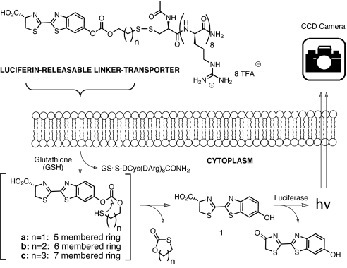
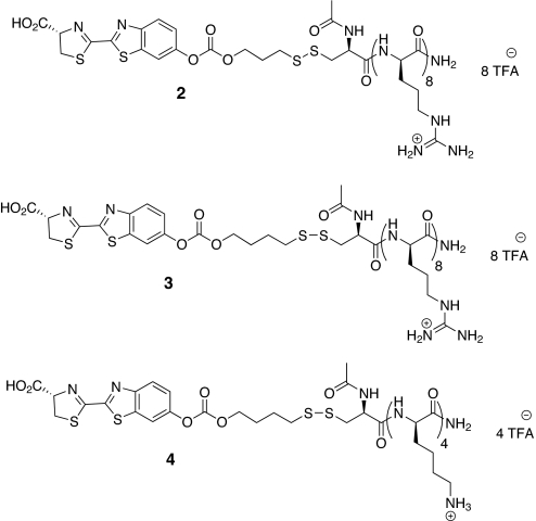
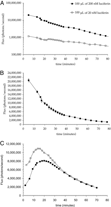
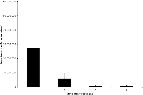
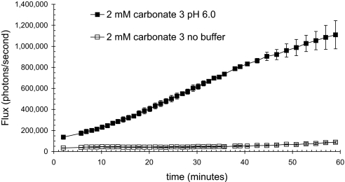
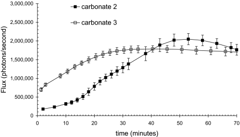
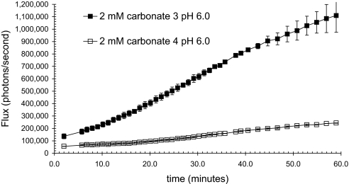
Many therapeutic leads fail to advance clinically because of bioavailability, selectivity, and formulation problems. Molecular transporters can be used to address these problems. Molecular transporter conjugates of otherwise poorly soluble or poorly bioavailable drugs or probes exhibit excellent solubility in water and biological fluids and at the same time an enhanced ability to enter tissues and cells and with modification to do so selectively. For many conjugates, however, it is necessary to release the drug/probe cargo from the transporter after uptake to achieve activity. Here, we describe an imaging method that provides quantification of transporter conjugate uptake and cargo release in real-time in animal models. This method uses transgenic (luciferase) reporter mice and whole-body imaging, allowing noninvasive quantification of transporter conjugate uptake and probe (luciferin) release in real time. This process effectively emulates drug-conjugate delivery, drug release, and drug turnover by an intracellular target, providing a facile method to evaluate comparative uptake of new transporters and efficacy and selectivity of linker release as required for fundamental studies and therapeutic applications.


Conflict of interest statement

The authors declare no conflict of interest.

Figures

Fig. 1.
Fig. 1.
Scheme for measurement of intracellular release.
Fig. 2.
Fig. 2.
Luciferin-releasable linker-transporter conjugates; conjuate 2, r8 transporter-disulfide linker-luciferin conjugate whose release would involve closure of a six-membered ring; conjugate 3, a homologous conjugate whose release would involve closure of a seven-membered ring; conjugate 4, negative control, analogous to 3, but incorporating a tetralysine, whose cellular uptake is minimal.
Fig. 3.
Fig. 3.
Resultant bioluminescence after intradermal injection of luciferin in HBS pH 7.4 into transgenic (FVB-luc+) mice. (A and B) Approximately 10 times the amount of light was observed when the luciferase expressing mice were injected with 200 vs. 20 nM luciferin (1) (A). Area under the curve for 200 nM was 3.02 × 10¹⁰ photons, whereas that for 20 nM was 3.11 × 10⁹ photons (10.24%). When plotted linearly, the bioluminescence rapidly decreases for the first 30 min (B). The plot is the average of three injections in separate animals. (C) Resultant bioluminescence after intradermal injection of 100 μl of 200 nM carbonate 2 in HBS pH 7.4 into luciferase transgenic (FVB-luc+) mice. The pattern of luminescence is shown for two different animals. The areas under the curve are 2.54 and 2.01 × 10¹⁰ photons.
Fig. 4.
Fig. 4.
Uptake of free luciferin (1) into skin immediately after depilatory treatment is significant and variable but is reduced with time to an insignificant level as the stratum corneum reestablishes itself. Total luminescence observed after topical application of 15 μl of 5.5 mM luciferin (1) in 200 mM NaOAc (pH 6.0) vehicle at various time points after treatment with Nair (Church and Dwight).
Fig. 5.
Fig. 5.
Inherent acidity of the trifluoroacetate salt of the conjugate 3 results in decreased bioluminescence. Differential bioluminescence observed when conjugate applied in buffered (filled squares) or unbuffered (open squares) vehicle. Carbonate 3 (2 mM) was applied in either 25% water/75% PEG 400 or 25% 200 mM NaOAc, pH 6.0/75% PEG 400.
Fig. 6.
Fig. 6.
Observed bioluminescence from luciferase transgenic mice as a function of time after topical application of 15 μl of 5 mM carbonate 2 and 3 in 75% PEG 400/25% 200 mM NaOAc, pH 6.0.
Fig. 7.
Fig. 7.
Observed bioluminescence from luciferase transgenic mice as a function of time after topical application of 15 μl of 2 mM carbonate 3 and 4 in 75% PEG 400/25% 1 M NaOAc, pH 6.0.

References

    1. Snyder EL, Dowdy SF. Pharm Res. 2004;21:389–393. - PubMed
    1. Trehin R, Merkle HP. Eur J Pharm Biopharm. 2004;58:209–223. - PubMed
    1. Fischer R, Fotin-Mleczek M, Hufnagel H, Brock R. ChemBioChem. 2005;6:2126–2142. - PubMed
    1. Gupta B, Levchenko TS, Torchilin VP. Adv Drug Deliv Rev. 2005;57:637–651. - PubMed
    1. Zorko M, Langel U. Adv Drug Deliv Rev. 2005;57:529–545. - PubMed

Publication types

LinkOut - more resources