. 2007 Jun 19;104(25):10601-6.
doi: 10.1073/pnas.0701096104. Epub 2007 Jun 11.

Attenuation of neuroinflammation and Alzheimer's disease pathology by liver x receptors

Affiliations

Attenuation of neuroinflammation and Alzheimer's disease pathology by liver x receptors

Noam Zelcer et al. Proc Natl Acad Sci U S A. .

Abstract

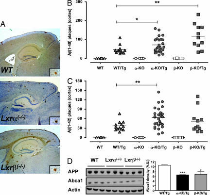
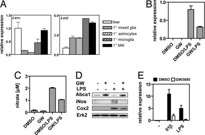
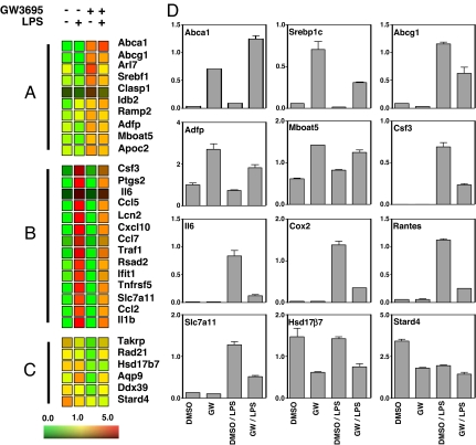
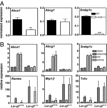
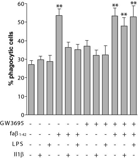
Alzheimer's disease (AD) is an age-dependent neurodegenerative disease that causes progressive cognitive impairment. The initiation and progression of AD has been linked to cholesterol metabolism and inflammation, processes that can be modulated by liver x receptors (LXRs). We show here that endogenous LXR signaling impacts the development of AD-related pathology. Genetic loss of either Lxralpha or Lxrbeta in APP/PS1 transgenic mice results in increased amyloid plaque load. LXRs regulate basal and inducible expression of key cholesterol homeostatic genes in the brain and act as potent inhibitors of inflammatory gene expression. Ligand activation of LXRs attenuates the inflammatory response of primary mixed glial cultures to fibrillar amyloid beta peptide (fAbeta) in a receptor-dependent manner. Furthermore, LXRs promote the capacity of microglia to maintain fAbeta-stimulated phagocytosis in the setting of inflammation. These results identify endogenous LXR signaling as an important determinant of AD pathogenesis in mice. We propose that LXRs may be tractable targets for the treatment of AD due to their ability to modulate both lipid metabolic and inflammatory gene expression in the brain.


Conflict of interest statement

The authors declare no conflict of interest.

Figures

Fig. 1.
Fig. 1.
Analysis of LXR-dependent gene expression in the brain. (A) LXRαβ−/− [knockout (KO)] and WT mice (n = six mice per group) were gavaged for 3 days with vehicle (Ve) or GW3695 (GW) (20 mg/kg). Brain RNA was analyzed individually for gene expression by quantitative PCR. Results are expressed as mean ± SD relative to vehicle-treated WT mice. (B) Immunoblot analysis of apoE in whole brain homogenates from male and female WT and LXRαβ−/− mice (n = two mice per group). The relative protein level was quantitated by densitometry.
Fig. 2.
Fig. 2.
Increased amyloid deposition in LXR-null mice. (A) Representative sagittal sections from 32-week-old WT/Tg, Lxrα−/−/Tg, and Lxrβ−/−/Tg mice stained for Aβ1–40. (Inset) A representative amyloid plaque. (Magnification: ×40.) (B and C) Amyloid Aβ1–40 (B) and Aβ1–42 (C) were quantified in 32-week-old mice. Each point represents an individual mouse, with the group average plotted as a bar. The number of mice and male/female distribution within the groups was WT (n = 6; 4/2), WT/Tg (n = 13; 9/4), α-KO (n = 4; 4/0), α-KO/Tg (n = 27; 15/12), β-KO (n = 4; 2/2), and β-KO/Tg (n = 11; 8/3). (D) Immunoblot analysis of APP and ABCA1 levels in whole-brain lysates of 12-week-old male mice of the indicated Tg genotype. Each lane represents an individual mouse. ∗, P < 0.05; ∗∗, P < 0.01.
Fig. 3.
Fig. 3.
Activated LXRs inhibit the inflammatory response of BV2 cells and primary microglia toward LPS and Il1β. (A) Relative expression of Lxrα and Lxrβ in primary mixed glia cultures, astrocytes, microglia, peritoneal macrophages, and liver. (B and C) BV2 cells were treated as indicated, and iNos expression (B) and nitrate release into the medium (C) were determined. (D) Immunoblot analysis of Abca1, iNos, and Cox2 levels in BV2 cells treated as indicated. Erk2 was used to control for loading. (E) Primary microglia were pretreated with DMSO or 1 μM GW3695 for 18 h and subsequently challenged with 15 ng/ml Il1β or 5 ng/ml LPS for 6 h, after which iNos expression was determined. Values shown are the average ± SD.
Fig. 4.
Fig. 4.
Transcriptional profiling of BV2 cells. BV2 cells were treated as indicated, and RNA was isolated and subjected to transcriptional profiling on Affymetrix arrays. (A–C) Heat diagrams of genes induced by GW3695 (A), genes induced by LPS (>5-fold) and repressed by GW3695 (>2-fold repression) (B), and genes repressed (>2-fold repression) by GW3695 (C). (D) Quantitative PCR analysis was used to confirm the changes in expression of the indicated genes in RNA samples from BV2 cells. Values shown are the average ± SD.
Fig. 5.
Fig. 5.
Analysis of gene expression in primary mixed glia. (A) Primary mixed glial cells were isolated from WT and Lxrαβ−/− mice, and the basal expression level of LXR target genes was analyzed. (B) Primary mixed glial cells were treated as indicated with DMSO or 1 μM GW3695 and in the presence or absence of 10 μM fAβ for 24 h. Transcript levels of the indicated genes were analyzed by quantitative PCR and are normalized to the DMSO-treated sample in each genotype. Values represent the mean ± SD of independent preparations made from WT (n = 4) and Lxrαβ−/− (n = 4) pups. ∗, P < 0.05.
Fig. 6.
Fig. 6.
LXRs regulate the phagocytic response of BV2 cells. BV2 cells were pretreated with DMSO or 1 μM GW3965 for 18 h and treated with or without 15 ng/ml Il1β or 100 ng/ml LPS for an additional 18 h. On the day of phagocytosis assay, the cells were treated with fAβ1–42 for 30 min and incubated with 1 μm of fluorescent microspheres for another 30 min. Phagocytosis of the fluorescent microspheres was quantified as percent of cell phagocytosing. Data represent the mean ± SE from three independent experiments. ∗∗, P < 0.001.

References

    1. Tontonoz P, Mangelsdorf DJ. Mol Endocrinol. 2003;17:985–993. - PubMed
    1. Zelcer N, Tontonoz P. J Clin Invest. 2006;116:607–614. - PMC - PubMed
    1. Janowski BA, Willy PJ, Devi TR, Falck JR, Mangelsdorf DJ. Nature. 1996;383:728–731. - PubMed
    1. Repa JJ, Mangelsdorf DJ. Annu Rev Cell Dev Biol. 2000;16:459–481. - PubMed
    1. Joseph SB, Castrillo A, Laffitte BA, Mangelsdorf DJ, Tontonoz P. Nat Med. 2003;9:213–219. - PubMed

Publication types

MeSH terms