. 2007 May 2:13:692-706.

Single-stranded oligonucleotide-mediated in vivo gene repair in the rd1 retina

Affiliations

Single-stranded oligonucleotide-mediated in vivo gene repair in the rd1 retina

Charlotte Andrieu-Soler et al. Mol Vis. .

Abstract

Purpose: The aim of this study was to test whether oligonucleotide-targeted gene repair can correct the point mutation in genomic DNA of PDE6b(rd1) (rd1) mouse retinas in vivo.

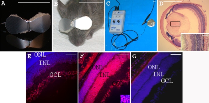
Methods: Oligonucleotides (ODNs) of 25 nucleotide length and complementary to genomic sequence subsuming the rd1 point mutation in the gene encoding the beta-subunit of rod photoreceptor cGMP-phosphodiesterase (beta-PDE), were synthesized with a wild type nucleotide base at the rd1 point mutation position. Control ODNs contained the same nucleotide bases as the wild type ODNs but with varying degrees of sequence mismatch. We previously developed a repeatable and relatively non-invasive technique to enhance ODN delivery to photoreceptor nuclei using transpalpebral iontophoresis prior to intravitreal ODN injection. Three such treatments were performed on C3H/henJ (rd1) mouse pups before postnatal day (PN) 9. Treatment outcomes were evaluated at PN28 or PN33, when retinal degeneration was nearly complete in the untreated rd1 mice. The effect of treatment on photoreceptor survival was evaluated by counting the number of nuclei of photoreceptor cells and by assessing rhodopsin immunohistochemistry on flat-mount retinas and sections. Gene repair in the retina was quantified by allele-specific real time PCR and by detection of beta-PDE-immunoreactive photoreceptors. Confirmatory experiments were conducted using independent rd1 colonies in separate laboratories. These experiments had an additional negative control ODN that contained the rd1 mutant nucleotide base at the rd1 point mutation site such that the sole difference between treatment with wild type and control ODN was the single base at the rd1 point mutation site.

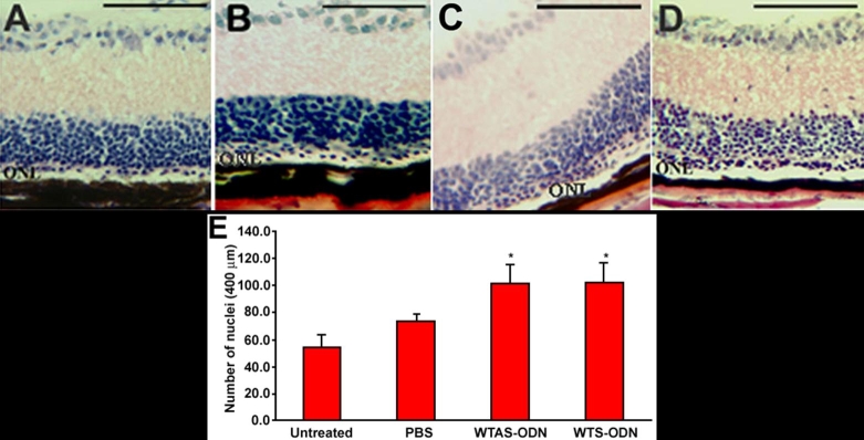
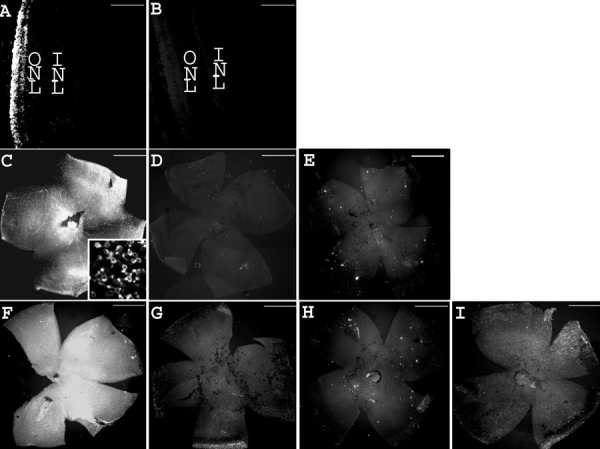
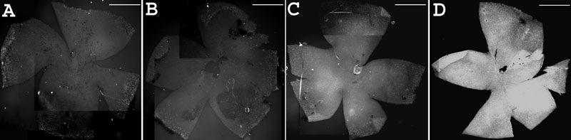
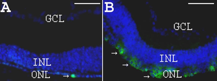
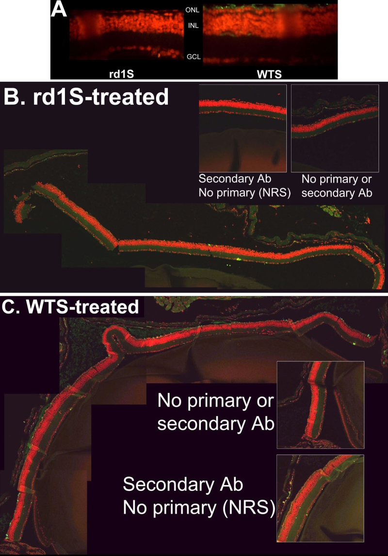
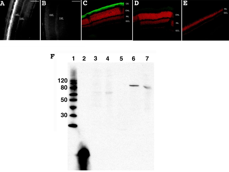
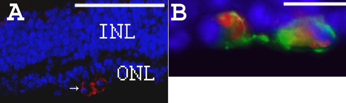
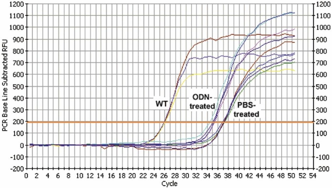
Results: Iontophoresis enhanced the penetration of intravitreally injected ODNs in all retinal layers. Using this delivery technique, significant survival of photoreceptors was observed in retinas from eyes treated with wild type ODNs but not control ODNs as demonstrated by cell counting and rhodopsin immunoreactivity at PN28. Beta-PDE immunoreactivity was present in retinas from eyes treated with wild type ODN but not from those treated with control ODNs. Gene correction demonstrated by allele-specific real time PCR and by counts of beta-PDE-immunoreactive cells was estimated at 0.2%. Independent confirmatory experiments showed that retinas from eyes treated with wild type ODN contained many more rhodopsin immunoreactive cells compared to retinas treated with control (rd1 sequence) ODN, even when harvested at PN33.

Conclusions: Short ODNs can be delivered with repeatable efficiency to mouse photoreceptor cells in vivo using a combination of intravitreal injection and iontophoresis. Delivery of therapeutic ODNs to rd1 mouse eyes resulted in genomic DNA conversion from mutant to wild type sequence, low but observable beta-PDE immunoreactivity, and preservation of rhodopsin immunopositive cells in the outer nuclear layer, suggesting that ODN-directed gene repair occurred and preserved rod photoreceptor cells. Effects were not seen in eyes treated with buffer or with ODNs having the rd1 mutant sequence, a definitive control for this therapeutic approach. Importantly, critical experiments were confirmed in two laboratories by several different researchers using independent mouse colonies and ODN preparations from separate sources. These findings suggest that targeted gene repair can be achieved in the retina following enhanced ODN delivery.


Figures

Figure 1
Figure 1
Iontophoresis device and eye sections from PN7 rd1 mice, 1 h after treatment. Iontophoresis device. A: An eye-glass-shaped electrode was made with aluminum foil and single-use disposable medical grade hydrophilic polyurethane sponge. B: The electrode covered both closed eyelids of the treated newborn mouse iontophoresis. C: shows the generator and the return electrode. Eye section 1 h after transpalpebral iontophoresis. D: Hematoxylin and eosin stained eye section showing integrity of the eye structures after iontophoresis Inset shows tissue at high magnification. Eye sections 1 h after intravitreal injection of CY3-tagged oligonucleotide. In E-G, nuclei were stained blue with DAPI and red with CY3. E: without prior iontophoresis, F: with prior iontophoresis (inset: high magnification of the ONL). G: Control retina from an rd1 mouse injected with 1 μl of PBS with prior iontophoresis. The following abbreviations were used: outer nuclear layer (ONL), inner nuclear layer (INL), and ganglion cell layer (GCL). Scale bars: A, B, C, 1 cm; D 1 mm; E, F, G and inset D, 100 μm; inset F, 5 μm.
Figure 2
Figure 2
Eye sections of treated and control PN28 rd1 mice and outer nuclear layer cell counting. Hematoxylin and eosin stained sections of rd1 eyes showed an increased number of nuclei rows in the outer nuclear layer (ONL) of oligonucleotide (ODN)-treated eyes. A: Untreated mouse. B: PBS-treated mouse. C: Mouse treated with WTAS ODN (corresponding to wild type antisense sequence). D: Mouse treated with WTS ODN (corresponding to wild type sense sequence). E: Counting of nuclei in the ONL shows a significant increase of nuclei in WTAS ODN- and WTS ODN-treated eyes compared to PBS-treated and untreated eyes (*p<0.05). Scale bars are A, B, C, and D, 100 μm.
Figure 3
Figure 3
Treatment with WTS preserves rhodopsin at PN28. Rhodopsin immunohistochemistry on wild type eye sections and rd1 whole flat-mount retinas, reflecting the time course of the retinal degeneration and the treatment efficacy. A: Wild type eye section from a mouse at PN28. B: Control eye section from wild type mouse at PN28 using normal mouse serum. C: rd1 flat-mount retina from a mouse at PN19 (inset: high magnification). D: Control flat-mount retina from rd1 mouse at PN19 using normal mouse serum. E: rd1 flat-mount retina from a mouse at PN28. F: PN28 rd1 flat-mount retina injected by WTS with prior iontophoresis at PN4, PN6, and PN8. G: PN28 rd1 flat-mount retina injected by WTS without prior iontophoresis at PN4, PN6, and PN8. H: PN28 rd1 flat-mount retina iontophoresed without oligonucleotide injection at PN4, PN6, and PN8. I: PN28 rd1 flat-mount retina injected with WTSscr7 with prior iontophoresis at PN4, PN6, and PN8. Scale bars are A and B, 100 μm; C, D, E, F, G, H, and I, 1 mm; inset, 10 μm.
Figure 4
Figure 4
Responsiveness of rhodopsin immunoreactivity to the number of oligonucleotide treatments. A: Three treatments with PBS (PN4, PN6, and PN8). B: One treatment with ODN at PN4. C: Two treatments with ODN (PN4 and PN6). D: Three treatments with ODN (PN4, PN6, and PN8). Scale bars : A, B, C, and D, 1 mm.
Figure 5
Figure 5
Rhodopsin immunohistochemistry on eye sections from PBS- or oligonucleotide-treated rd1 mice at PN28. A: DAPI staining in blue and rho-4D2 immunostaining in green (arrows) on section from PN28 PBS-treated rd1 retina. B: DAPI staining in blue and rho-4D2 immunostaining in green (arrows) on section from PN28 ODN-treated rd1 retina. Scale bars are A and B, 150 μm.
Figure 6
Figure 6
Effect of treatment with WTS versus rd1S at PN33: A confirmatory experiment. In experiments conducted with an independent C3H/henJ colony, littermates treated with WTS ODN had many more rhodopsin-immunopositive cells in the ONL than littermates treated with rd1S, the identical ODN with the exception of having the mutant rd1 nucleotide at the rd1 mutation site. Tissue was harvested and sections prepared at PN33. A: Fluorescent micrographs of retina sections from rd1S-treated mouse (left panel) and WTS-treated mouse (right panel). Rhodopsin immunosignal is green, counterstained nuclei are red. B: Composite image of confocal micrographs of retina section from mouse treated with rd1S. Rhodopsin immunosignal is green, counterstained nuclei are red. Very little rhodopsin signal is apparent. Insets are control sections in which no antibody was used or secondary antibody was used by normal rabbit sera (NRS) was substituted for primary antibody. C: Composite image of confocal micrographs of retina section from mouse treated with WTS. Many rhodopsin-positive cells are apparent in the putative photoreceptor layer. Insets are as in B. Quantification of rhodopsin-positive cells in photoreceptor layers showed that significantly more signal was observed with WTS treatment compared to rd1S treatment (see "Results").
Figure 7
Figure 7
β-PDE immunohistochemistry and western blot. A: Wild type +/+ eye section from a mouse at PN28. B: Control wild type +/+ eye section from a mouse at PN28 using normal rabbit sera. C: Confocal micrograph from a C57/BL6 (wild type; +/+) retina reacted with β-PDE primary antibody and Oregon Green-conjugated goat anti-rabbit IgG (giving green signal in figure) and counter-stained with propidium iodide (giving red signal in figure). D: Confocal micrograph from wild type retina reacted with normal rabbit serum. E: Confocal micrograph from a C3H/henJ mouse (rd1; -/-) retina reacted with β-PDE primary antibody and Oregon green-conjugated goat anti-rabbit IgG. OS indicates outer segments, ONL indicates outer nuclear layer, INL indicates inner nuclear layer, GCL indicates ganglion cell layer. F: Anti-β-PDE "western" immunoblot. Lane 1 is the molecular weight marker (sizes given on left), lane 2 is the polypeptide antigen against which the antibody was raised, lane 3 is protein from a C3H (rd1) mouse retina, lane 4 is protein from a rd10 mouse retina, lane 5 is protein from an FVB (rd1) mouse retina, lane 6 is protein from a CCRC (wild type +/+) mouse retina, lane 7 is protein from a Balb/C (wild type +/+) mouse retina. All mice were at least 30 days old at time of tissue harvest. Scale bars are A, and B, 100 μm. These data suggest that the antibodies are highly specific and selective to β-PDE.
Figure 8
Figure 8
Rhodopsin and β-PDE immunohistochemistry on eye sections from PBS- or WTS ODN-treated rd1 mice at PN28. A: DAPI staining in blue and β-PDE immunostaining in red (arrows) on section from PN28 ODN-treated rd1 retina. B: Combined fluorescence of β-PDE immunostaining in red, rho-4D2 immunostaining in green and DAPI staining in blue on section from PN28 ODN-treated rd1 retina. Scale bars are A, 150 μm; B, 10 μm. β-PDE immunoreactivity is associated with cells in the remaining outer nuclear layer (ONL). This staining appears associated with the cytoplasm, not the nucleus. The rhodopsin immunoreactivity is associated with the same cells in the ONL and the immunoreactivity seems more peripheral to the cytosolic β-PDE staining, suggesting a plasma membrane localization of the rhodopsin immunoreactivity. Relatively few cells in the residual ONL exhibit either rhodopsin or β-PDE immunoreactivity at P28 even after three ODN treatments; however, those remaining positive cells exhibit some degree of clustering.
Figure 9
Figure 9
Representative plots of allele specific real time polymerase chain reaction. The graph shows the real-time detection of fluorescence resulting from intercalation of SybrGreen fluorescent dye into double-stranded PCR products. Template DNA was isolated from BALB/c mouse (WT), retinas of rd1 mice treated with WTS oligonucleotide (ODN-treated), or retinas of rd1 mice treated with PBS (PBS-treated). Primers were specific for wild type allele. Each curve represents a different eye. Each experimental sample was assayed in 5-10 replicates. See "Materials and Methods" for details of detection threshold calculation (orange horizontal line), normalization, and quantification. Control experiments in which WT ODNs were doped into rd1 DNA show no shift to the left. Numerous experiments with rd1 DNAs isolated from many different individual mouse eyes all showed the typical curve of the PBS-treated samples crossing threshold at about 37 cycles.

References

    1. Farber DB, Lolley RN. Cyclic guanosine monophosphate: elevation in degenerating photoreceptor cells of the C3H mouse retina. Science. 1974;186:449–51. - PubMed
    1. Yan W, Lewin A, Hauswirth W. Selective degradation of nonsense beta-phosphodiesterase mRNA in the heterozygous rd mouse. Invest Ophthalmol Vis Sci. 1998;39:2529–36. - PubMed
    1. Farber DB, Danciger M. Identification of genes causing photoreceptor degenerations leading to blindness. Curr Opin Neurobiol. 1997;7:666–73. - PubMed
    1. Pittler SJ, Baehr W. Identification of a nonsense mutation in the rod photoreceptor cGMP phosphodiesterase beta-subunit gene of the rd mouse. Proc Natl Acad Sci USA. 1991;88:8322–6. - PMC - PubMed
    1. McLaughlin ME, Ehrhart TL, Berson EL, Dryja TP. Mutation spectrum of the gene encoding the beta subunit of rod phosphodiesterase among patients with autosomal recessive retinitis pigmentosa. Proc Natl Acad Sci USA. 1995;92:3249–53. - PMC - PubMed

Publication types

MeSH terms

LinkOut - more resources