Review
. 2007 Jul;8(7):555-63.
doi: 10.1038/nrg2125. Epub 2007 Jun 12.

Planar cell polarity: one or two pathways?

Affiliations
Review

Planar cell polarity: one or two pathways?

Peter A Lawrence et al. Nat Rev Genet. 2007 Jul.

Abstract

In multicellular organisms, cells are polarized in the plane of the epithelial sheet, revealed in some cell types by oriented hairs or cilia. Many of the underlying genes have been identified in Drosophila melanogaster and are conserved in vertebrates. Here we dissect the logic of planar cell polarity (PCP). We review studies of genetic mosaics in adult flies - marked cells of different genotypes help us to understand how polarizing information is generated and how it passes from one cell to another. We argue that the prevailing opinion that planar polarity depends on a single genetic pathway is wrong and conclude that there are (at least) two independently acting processes. This conclusion has major consequences for the PCP field.


Figures
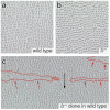
Figure 1
Figure 1. Planar cell polarity in the Drosophila pleura
a. In the wildtype, the small cuticular hairs (several are produced by each cell) point posteriorly. b. In fz flies, the orientation is randomised. c. Three clones of fz cells in a wildtype fly, each outlined in red; the hairs posterior to the fz cells mostly point anteriorwards (red arrows) but in the strait between two nearby clones, polarity is normal (black arrow) The fz cells (one is filled in orange to show its extent) are genetically marked so that each makes numerous hairs that point upwards.
Figure 2
Figure 2. The functional assay
Clones (blank) are made in the Drosophila abdomen of one genotype (the “sending” cells) that may alter the polarity of hairs in the “receiving” cells (grey background). Anterior to the top. Black arrows, normal polarity; blue arrows, disturbed; red arrows, reversed polarity. a. fz cells reverse the polarity of receiving cells posterior to the clone, while overexpression of fz (UAS.fz) reverses the polarity anterior to the clone. The assays show that Stan is needed in both sending and receiving cells. b. The signal emanating from UAS.ft cells acts independently of Stan. c. These three assays argue that Fj acts through Ft. UAS.fj clones reverse receiving cells in front, like UAS.ft clones (d.) but if Ft is missing, as in a UAS.fj, ft clone, it behaves like a ft clone (which reverse receiving cells behind the clone). d. These 7 assays plus the ft clones (c.) show that Ds alone is sufficient in the sending cells to affect polarity of the receiving cells (similar results show that Ft alone is also sufficient32), but that both Ds and Ft are needed in the receiving cells.
Figure 3
Figure 3. A repolarising clone in a stan fly
The clone is illustrated as in Fig 1, and consists of marked cells expressing a modified form of Ds in a fly mutant for the stan gene. In spite of the lack of the Stan system, the clone reverses the polarity of cells behind it, and also organises normal polarity in the cells in front of it (a clone of the same genotype is shown in Fig 2F of 32)
Figure 4
Figure 4. Cuticle from the dorsal abdomen of Drosophila
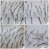
a. wildtype cuticle b. dsc. stand. ds stan. Note the stronger randomisation of hairs and bristles when both systems are broken.
Figure 5
Figure 5. The localisation of PCP proteins in clones in the wing
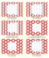
The proteins may be located on particular faces of the cell membranes (protein shown in red). Anterior to the top, distal to the right. a. Stan accumulates mostly on the proximodistal faces of the cells and is not seen at all in the membrane unless Stan protein is present on both confronting cells. b. Fz protein is also seen mostly on the proximodistal faces of the cells and, if tagged with GFP, the clone shows that Fz does not go to the membrane without the Stan protein. c. Fz is actually localised on the distal face of the cells; the white cells are a clone of cells that lack the tagged Fz. d. In cells lacking Pk, Fz accumulates uniformly, with no asymmetry. e, f. Evidence for Ds-Ft heterodimers. A comparison of both cases shows that Ds protein only accumulates at the membrane when there is Ft protein on the facing membrane of the neighbouring cell.
Figure 6
Figure 6. Alternative models of PCP
a. The single pathway model. Morphogens drive gradients of Fj and Ds expression that affects the Ds system by a small differential input into the proximal and distal faces of the membrane of each cell. This would influence, somehow, the distribution of the Dsh, Pk, Vang and Fz in the membrane which, through a feedback loop, would be amplified to polarise the cells, . b. The two pathway model. On the left, the Ds system is shown; here the morphogens drive gradients of Fj and Ds expression so that both Ft and Ds become graded across the field of cells as a gradient of Ds-Ft heterodimers. The difference between the numbers of these on the anterior and posterior faces might polarise each cell. The evidence for this model comes from the functional assays, see text. On the right, the Stan system is shown. Here the morphogens might feed directly into Fz, setting up a Fz activity gradient across the whole field of cells. Using Stan, the level of Fz activity of neighbouring cells is compared, so that each cell becomes oriented to point towards the neighbour with the lowest Fz activity.

References

    1. Brenner S. My Life in Science. BioMed Central; 2001.
    1. Drubin D. In: Cell Polarity. Hames B, Glover D, editors. Oxford University Press; Oxford: 2000.
    1. Gho M, Schweisguth F. Frizzled signalling controls orientation of asymmetric sense organ precursor cell divisions in Drosophila. Nature. 1998;393:178–81. - PubMed
    1. Lawrence PA. Development and determination of hairs and bristles in the milkweed bug, Oncopeltus fasciatus (Lygaeidae, Hemiptera) J. Cell Science. 1966;1:475–498. - PubMed
    1. Wang Y, Thekdi N, Smallwood PM, Macke JP, Nathans J. Frizzled-3 is required for the development of major fiber tracts in the rostral CNS. J Neurosci. 2002;22:8563–73. - PMC - PubMed

Publication types

LinkOut - more resources