. 2007 Jul;81(1):1-16.
doi: 10.1086/518428. Epub 2007 May 15.

Mutations in FGD4 encoding the Rho GDP/GTP exchange factor FRABIN cause autosomal recessive Charcot-Marie-Tooth type 4H

Affiliations

Mutations in FGD4 encoding the Rho GDP/GTP exchange factor FRABIN cause autosomal recessive Charcot-Marie-Tooth type 4H

Valérie Delague et al. Am J Hum Genet. 2007 Jul.

Abstract

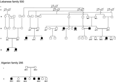
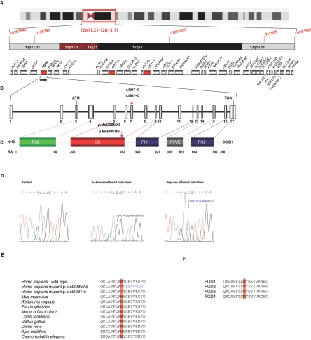
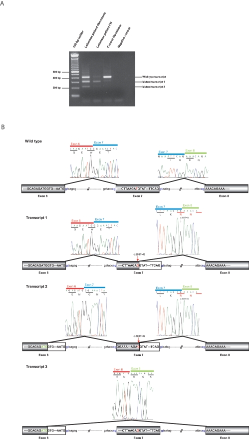
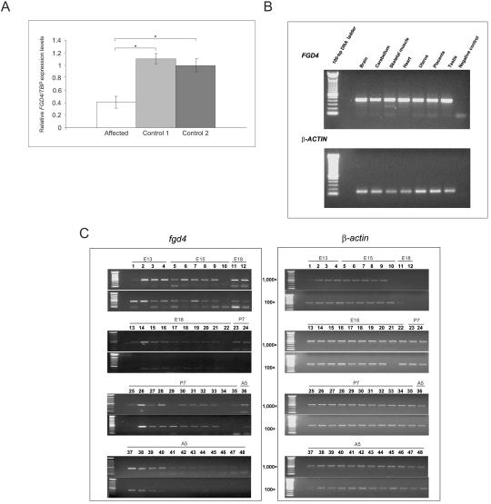
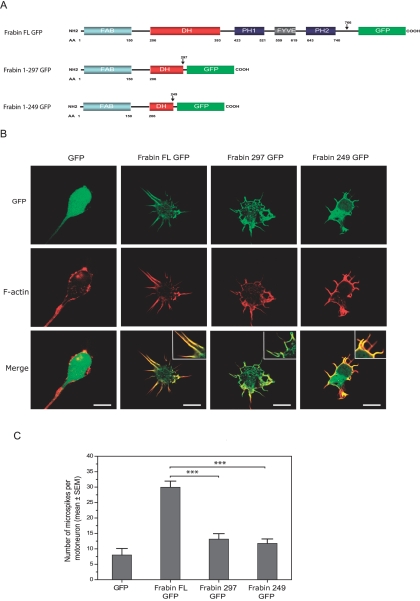
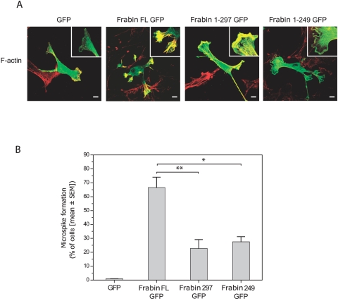
Charcot-Marie-Tooth (CMT) disorders are a clinically and genetically heterogeneous group of hereditary motor and sensory neuropathies characterized by muscle weakness and wasting, foot and hand deformities, and electrophysiological changes. The CMT4H subtype is an autosomal recessive demyelinating form of CMT that was recently mapped to a 15.8-Mb region at chromosome 12p11.21-q13.11, in two consanguineous families of Mediterranean origin, by homozygosity mapping. We report here the identification of mutations in FGD4, encoding FGD4 or FRABIN (FGD1-related F-actin binding protein), in both families. FRABIN is a GDP/GTP nucleotide exchange factor (GEF), specific to Cdc42, a member of the Rho family of small guanosine triphosphate (GTP)-binding proteins (Rho GTPases). Rho GTPases play a key role in regulating signal-transduction pathways in eukaryotes. In particular, they have a pivotal role in mediating actin cytoskeleton changes during cell migration, morphogenesis, polarization, and division. Consistent with these reported functions, expression of truncated FRABIN mutants in rat primary motoneurons and rat Schwann cells induced significantly fewer microspikes than expression of wild-type FRABIN. To our knowledge, this is the first report of mutations in a Rho GEF protein being involved in CMT.


Figures

Figure  1.
Figure 1.
Pedigrees of the Lebanese (500) and Algerian (295) families. Blackened symbols indicate affected individuals.
Figure  2.
Figure 2.
CMT4H locus, structure of FGD4 and FRABIN, mutations, and conservation between species. A, Chromosomal region at 12p11.21-q13.11 containing the CMT4H candidate locus, as identified elsewhere, covering a 15.8-Mb region located between STR markers D12S1648 and D12S1661. STR markers homozygous by descent are shown in red. The 48 known genes lying in this region are schematically represented, with candidate genes tested in our study indicated in red (FGD4, CNTN1, KIF21A, and RAPGEF3). B, Structure of FGD4. The gene is located at 12p11.21, near the centromere, and is transcribed from telomere to centromere, as indicated by the black arrow. The gene, covering a genomic region of 138,580 bp, as defined by the GenBank reference sequence NM_139241, is composed of 17 exons. The transcript is 2,931 bp long, with a coding sequence (exons 3–17) of 2,301 bp (for mRNA [GenBank accession number AK057294]). The coding region is indicated in gray and UTRs (5′ and 3′) in white. The coding sequence includes exons 3–17. Additional exons found in alternative splice variants are described in the databases but are not represented here. C, Five functional domains presented by FRABIN, encoded by FGD4: FAB, DH presenting a Rho GEF activity, PH1 and PH2, which bind to phosphatidylinositol(3,4,5)P3, and one FYVE domain that interacts with phosphatidylinositol3P. D, Chromatograms showing the two mutations identified in FGD4: the c.893T→G transversion in the Lebanese patients and the c.893T→C transition in the Algerian patient. E and F, The methionine at residue 298, mutated in both patients, which is highly conserved in different species (vertebrates and nonvertebrates), as well as in other members of the human FGD family.
Figure  3.
Figure 3.
The c.893T→G splicing mutation. A, Results of RT-PCR experiments performed on the Lebanese patient’s fibroblasts and peripheral nerve (PN) and in the control fibroblasts, with use of a forward primer located in exon 5 and a reverse primer located in exon 8 (see table 1). In control fibroblasts, a 412-bp band corresponding to the wild-type transcript was obtained. In the Lebanese patient’s fibroblasts, we observed two additional bands corresponding to transcripts 1 and 2 described in panel B. In fibroblasts, a band the size of the wild-type transcript was observed, corresponding to the correctly spliced transcript with a homozygous p.Met298Arg missense mutation. B, Schematic representation of splicing defects caused by the c.893T→G Lebanese mutation observed in panel A. In the first mutant (transcript 1), the GT donor splice site created by the mutation was used instead of the usual consensus 3′ donor site. The transcript deleted from the last 100 bp of exon 7 is predicted to yield a 305-aa truncated protein, and the mutation is described as “p.Met298fsX8.” In transcript 2 (224 bp), in addition to the last 100 bp of exon 7, the last 99 bp of exon 6 are deleted because of the use of a cryptic donor splice site in exon 6. Transcript 3, identified after the cloning of RT-PCR fragments obtained from the patient’s lymphoblast cDNA, lacks the last 99 bp of exon 6 and all of exon 7.
Figure  4.
Figure 4.
Expression analysis. A, RTQ-PCR experiments with use of cDNAs extracted from the patient’s fibroblasts and two controls. TBP was used as the reference gene for normalization. An approximate average twofold underexpression of FGD4 was observed in Lebanese patients with CMT4H as compared with calibrators (P<.05, Wilcoxon-Mann-Whitney test). Similar results were obtained using normalization with GUSB (data not shown). B, Semiquantitative expression of FGD4 in different human adult tissues. β-actin was used for normalization. C, Expression of FGD4 in different mouse nerve tissues. Two cDNA concentrations were used (100× and 1,000×). β-actin was used for normalization. Lanes (L) correspond to the following: L1, embryo day 13 (E13) telencephalon/diencephalons. L2, E13 mesencephalon (midbrain). L3, E13 rhombencephalon (hindbrain). L4, E13 spinal cord. L5, E15 telencephalon. L6, E15 diencephalon. L7, E15 midbrain. L8, E15 pons. L9, E15 medulla. L10, E15 spinal cord. L11, E18 frontal cortex. L12, E18 posterior cortex. L13, E18 entorhinal cortex. L14, E18 olfactory bulb. L15, E18 hippocampus. L16, E18 striatum. L17, E18 thalamus. L18, E18 hypothalamus. L19, E18 midbrain. L20, E18 pons. L21, E18 medulla. L22, E18 spinal cord. L23, postnatal day 7 (P7) frontal cortex. L24, P7 posterior cortex. L25, P7 entorhinal cortex. L26, P7 olfactory bulb. L27, P7 hippocampus. L28, P7 striatum. L29, P7 thalamus. L30, P7 hypothalamus. L31, P7 cerebellum. L32, P7 midbrain. L33, P7 pons. L34, P7 medulla. L35, P7 spinal cord. L36, adult 5-wk (A5) frontal cortex. L37, A5 posterior cortex. L38, A5 entorhinal cortex. L39, A5 olfactory bulb. L40, A5 hippocampus. L41, A5 striatum. L42, A5 thalamus. L43, A5 hypothalamus. L44, A5 cerebellum. L45, A5 midbrain. L46, A5 pons. L47, A5 medulla. L48, A5 spinal cord. FGD4 was expressed in all parts of the brain and in spinal cord, but with stronger expression at embryonic and prenatal stages than at postnatal and adult stages.
Figure  5.
Figure 5.
A, Domain organization of rat Frabin constructs. Frabin FL GFP = full-length Frabin C-terminally tagged to GFP. Frabin 1–297 GFP = GFP-tagged Frabin lacking the last 469 aa, mimicking the predicted protein resulting from mutant transcript 1 (see fig. 3); Frabin 1–249 GFP = GFP-tagged Frabin lacking the last 513 aa, mimicking the protein corresponding to mutant transcript 2 (see fig. 3). B, Confocal images of rat motoneurons transduced with GFP or GFP-tagged Frabin expression constructs and labeled at 1 DIV for f-actin using Alexa Fluor 546-conjugated phalloidin. While GFP shows uniform cytoplasmic expression, FL Frabin GFP colocalizes with f-actin in microspikes and induces their formation (see merged images and insets). Note reduced number and abnormal shape of microspikes induced by expression of truncated Frabin constructs. Scale bar = 10 μm. C, Diagram showing reduced microspike formation in motoneurons expressing truncated forms of Frabin as compared with motoneurons expressing full-length Frabin (n=25 cells per condition, P<.001 as assessed by Mann-Whitney rank sum test).
Figure  6.
Figure 6.
A, Confocal images showing cultured rat RT4 schwann cells transfected with expression constructs for GFP, full-length (FL) Frabin-GFP or truncated Frabin-GFP (see fig. 5). Cells are co-labeled for f-actin (in red); details of membrane structures are magnified (see insets). Scale bars = 10 μm. B, Diagram showing reduced microspike formation in RT4 cell cultures transfected with truncated forms of Frabin (Frabin 297 GFP and Frabin 249 GFP) as compared with cultures transfected with Frabin FL GFP (* indicates P=.03 and ** indicates P=.011, as assessed by Student t test).
Figure  7.
Figure 7.
Labeling of microfilament and microtubule cytoskeleton and centrosomes in the control and Lebanese patient (500.21) fibroblasts (see fig. 1). A, Labeling of microfilament (antibody to F-actin) and microtubules (antibody to α-tubulin). No significant differences were observed between the control and patient fibroblasts. B, Labeling of total microtubules (antibody to α-tubulin) and stable microtubules (antibody to acetylated α-tubulin). Stabilized microtubules were denser at centrosomes, but no structural difference was noticed in patients compared with controls. C, Labeling of centrosome with use of antibody to γ-tubulin or pericentrin and antibody to α-tubulin. Centrosome position and structure seemed normal in both patient and control fibroblasts.

References

Web Resources

    1. GenBank, http://www.ncbi.nlm.nih/Genbank/ (for KIF21A [accession number AY368076]; RAPGEF3 [accession number U78168]; FGD4 [accession number NM_139241]; FGD4 mRNA [accession numbers AK057294, BC045552, and AY367054] and ESTs for H. sapiens [accession number AA305646] and Macaca mulatta [accession number CN643653]; Fgd4 [accession number NM_139234]; FGD4 homologs FGD1 [accession number NP_004454], FGD2 [accession number NP_775829], and FGD3 [NP_149077]; and FRABIN proteins for H. sapiens [accession number NP_640334], Rattus norvegicus [accession number NP_640356], Mus musculus [accession number NP_631978], Macaca fascicularis [accession number BAE90450], Caenorhabditis elegans [accession number NP_001023039], Canis familiaris [accession number XP_543741], Pan troglodytes [accession number XP_520721], Gallus gallus [accession number XP_416365], Apis mellifera [accession number XP_394280], and Danio rerio [accession number CAK11116])
    1. Inherited Peripheral Neuropathies Mutation Database, http://www.molgen.ua.ac.be/CMTMutations/default.cfm
    1. Neuromuscular Disease Center, http://www.neuro.wustl.edu/neuromuscular/time/hmsn.html (for comprehensive and updated data about CMT diseases)
    1. Online Mendelian Inheritance in Man (OMIM), http://www.ncbi.nlm.nih.gov/Omim/ (for GDAP1, CMT4A, MTMR2, CMT4B1, SBF2/MTMR13, CMT4B2, SH3CT2/KIAA1985, CMT4C, NDRG1, HMSN-Lom, EGR2, P0, PMP22, CMT4E, Dejerine-Sottas syndrome, PRX, CMT4F, HMSN-Russe, and CMT4H)
    1. ORF Finder, http://www.ncbi.nlm.nih.gov/gorf/gorf.html

References

    1. Skre H (1974) Genetic and clinical aspects of Charcot-Marie-Tooth’s disease. Clin Genet 6:98–118 - PubMed
    1. Dyck PJ, Chance P, Lebo R, Carney AJ (1993) Hereditary motor and sensory neuropathies. In: Dyck PJ, Thomas PK, Griffin JW, Low PA, Podulso JF (eds) Peripheral neuropathy. WB Saunders, Philadelphia, pp 1094–1136
    1. Villanova M, Timmerman V, De Jonghe P, Malandrini A, Rizzuto N, Van Broeckhoven C, Guazzi G, Rossi A (1998) Charcot-Marie-Tooth disease: an intermediate form. Neuromuscul Disord 8:392–39310.1016/S0960-8966(98)00044-3 - DOI - PubMed
    1. Shy ME (2004) Charcot-Marie-Tooth disease: an update. Curr Opin Neurol 17:579–58510.1097/00019052-200410000-00008 - DOI - PubMed
    1. Berciano J, Combarros O (2003) Hereditary neuropathies. Curr Opin Neurol 16:613–62210.1097/00019052-200310000-00008 - DOI - PubMed

Publication types

MeSH terms

Substances