. 2007 Jul;81(1):67-76.
doi: 10.1086/518900. Epub 2007 May 16.

The nuclear factor kappaB-activator gene PLEKHG5 is mutated in a form of autosomal recessive lower motor neuron disease with childhood onset

Affiliations

The nuclear factor kappaB-activator gene PLEKHG5 is mutated in a form of autosomal recessive lower motor neuron disease with childhood onset

Isabelle Maystadt et al. Am J Hum Genet. 2007 Jul.

Abstract

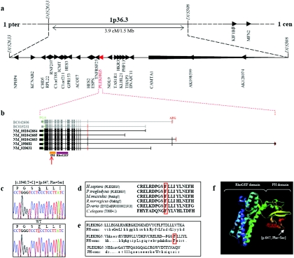
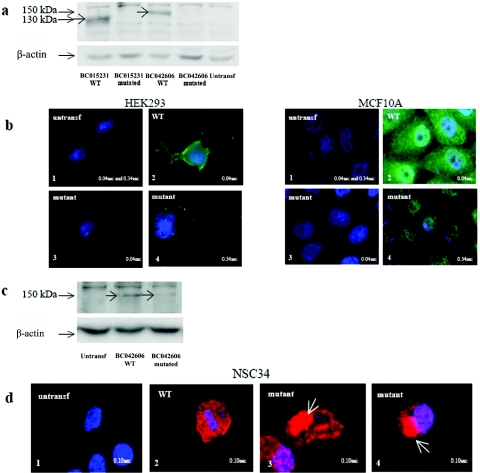
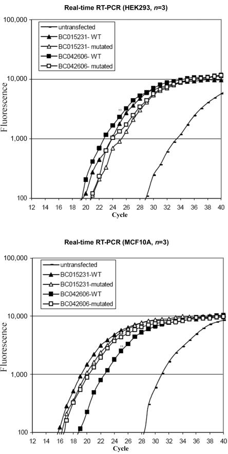
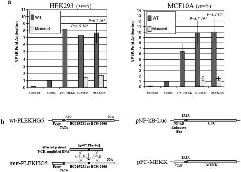
Lower motor neuron diseases (LMNDs) include a large spectrum of clinically and genetically heterogeneous disorders. Studying a large inbred African family, we recently described a novel autosomal recessive LMND variant characterized by childhood onset, generalized muscle involvement, and severe outcome, and we mapped the disease gene to a 3.9-cM interval on chromosome 1p36. We identified a homozygous missense mutation (c.1940 T-->C [p.647 Phe-->Ser]) of the Pleckstrin homology domain-containing, family G member 5 gene, PLEKHG5. In transiently transfected HEK293 and MCF10A cell lines, we found that wild-type PLEKHG5 activated the nuclear factor kappa B (NF kappa B) signaling pathway and that both the stability and the intracellular location of mutant PLEKHG5 protein were altered, severely impairing the NF kappa B transduction pathway. Moreover, aggregates were observed in transiently transfected NSC34 murine motor neurons overexpressing the mutant PLEKHG5 protein. Both loss of PLEKHG5 function and aggregate formation may contribute to neurotoxicity in this novel form of LMND.


Figures

Figure  1.
Figure 1.
Physical map of the locus associated with generalized LMND, structure of PLEKHG5, and location of the mutation identified in the African family. a, Location and transcriptional direction of the 27 sequenced genes (UCSC Genome Browser). b, Structure of PLEKHG5 (UCSC Genome Browser). The five isoforms annotated by NCBI genome browser are in black, and the two Mammalian Gene Collection Full ORF mRNAs used for plasmid constructions are in gray. The coding exons are shown as vertical bars, illustrating their approximate size and position. The position of the initiation codon (ATG) at nucleotide +1 is indicated in red, and the position of the stop codon (TGA) is indicated in green. The 5′ and 3′ UTRs are shown as shorter vertical bars. Locations of the PH and RhoGEF domains are also shown (Pfam). The identified homozygous mutation is located in the PH domain (red arrow). c, Electrophoregram of affected individuals compared with a control, showing the homozygous c.1940 T→C (p.647 Phe→Ser) mutation (GenBank accession number NM_020631.2). WT = wild type. d, Phenylalaline at position 647 (boxed in red) highly conserved across species: Homo sapiens, Pan troglodytes, Mus musculus, Rattus norvegicus, Danio rerio, and Caenorhabditis elegans (UCSC Genome Browser). e, Comparison of the PH domain of PLEKHG5 with the PH domain consensus sequence (PH-cons). In this sequence, the eight consensus residues are indicated in capital letters, including the mutant phenylalanine (boxed in red) (Pfam). f, Predictive three-dimensional structure of PLEKHG5 (ModBase). The RhoGEF domain has an α-helical structure, and the PH domain consists of seven β-sheets, followed by a C-terminal amphipathic helix. The mutation occurs in the area of the β5/β6 loop of the PH domain, which may be part of the phospholipids binding site.
Figure  2.
Figure 2.
Expression of PLEKHG5 in the human nervous system. PLEKHG5 expression was evaluated in several human nervous tissues by real-time quantitative RT-PCR. All reactions were performed in triplicate, and PLEKHG5 expression was normalized according to β2 microglobulin expression. Results were expressed as amplification rates in comparison with PLEKHG5 expression in untransfected HEK293 cell line. PLEKHG5 transcripts were detected in all studied tissues, supporting its ubiquitous expression pattern in the human nervous system, with a predominance in peripheral nerve and spinal cord. Sm = skeleton muscle; Br = brain; Sc = spinal cord; Pn = peripheral nerve.
Figure  3.
Figure 3.
Analysis of wild-type (WT) and mutant PLEKHG5 protein expression in untransfected (untransf) and transiently transfected HEK293, MCF10A, and NSC34 cells. a, Analysis by western blotting of PLEKHG5 protein expression in HEK293 cells. PLEKHG5 protein was not detectable in untransfected cells. The 130-kDa and 150-kDa bands were detected in cells overexpressing the BC015231 and BC042606 wild-type isoforms. In contrast, no similar bands were observed in cells transfected with the mutant variants. Similar results were found after distinct protein extraction methods (with 0.32 M sucrose or lysis buffer A; see the “Subjects and Methods” section) and in both HEK293 and MCF10A cells. b, Immunofluorescence assays of PLEKHG5 protein expression in HEK293 and MCF10A cells. b1, There was an absence of signal in untransfected cells, even with a long exposure time (0.34 s). b2, With an exposure time of 0.04 s, cells transfected with wild-type PLEKHG5 showed a diffuse cytoplasmic distribution of PLEKHG5 protein. b3, In cells transfected with the mutant constructs, no protein was visualized with an identical exposure time (0.04 s). b4, Increased exposure time (0.34 s) revealed a weak signal in the nucleus and the cytoplasm. c, Analysis by western blotting of PLEKHG5 protein expression in NSC34 cells. Wild-type PLEKHG5 protein was not detectable in untransfected cells. The 150-kDa band was detected in cells overexpressing the BC042606 wild-type isoform. In contrast, the band intensity was clearly decreased in cells transfected with the BC042606 mutant variant. d, Immunofluorescence assays of PLEKHG5 protein expression in NSC34 cells. The transfection efficacy was controlled using cotransfection with a GFP-expressing plasmid. d1, There was an absence of signal in GFP-negative nontransfected cells. d2, Cells transfected with the wild-type PLEKHG5 plasmid showed a diffuse cytoplasmic distribution of the PLEKHG5 protein. d3 and d4, Presence of large immunoreactive aggregates (white arrows) in the soma of NSC34 motor neurons transfected with the mutant PLEKHG5 plasmids.
Figure  4.
Figure 4.
Quantification, by real-time RT-PCR, of PLEKHG5 expression levels in untransfected and transfected HEK293 and MCF10A cells. At 48 h after transfection, the cells were harvested and were analyzed by real-time RT-PCR for PLEKHG5 mRNA. The fluorescence was measured in every PCR cycle and was proportional to the accumulation of PCR product. Endogenous expression of PLEKHG5 was detected in untransfected control cells but was markedly increased in transfected cells (up to 1,000-fold, estimated by ΔΔCt method). Overexpression of wild-type (WT) and mutated PLEKHG5 in transfected cells was similar. Triplicate analyses were performed for each sample.
Figure  5.
Figure 5.
Comparison between wild-type (WT) and mutated PLEKHG5 activity on the NFκB signaling pathway. a, Reporter construct consisting of the luciferase gene placed under control of a promoter containing the consensus NFκB binding sites (Stratagene), transfected in HEK293 or MCF10A cells alone (Control), in combination with expression vectors for wild-type and mutated PLEKHG5 proteins (BC015231 and BC042606), or in combination with expression vector for a positive control of NFκB activation (pFC-MEKK). Values are expressed as fold activation over transfection of the reporter plasmid alone. Bars indicate the SD of five independent experiments. The differences between wild-type and mutated PLEKHG5 are statistically significant according to Welch’s t test. b, Schematic representation of the plasmid constructions (see the “Subjects and Methods” section). Untransf. = untransfected. PCMV = CMV promoter; TATA = TATA box.

References

Web Resources

    1. Expression Profile Viewer, http://www.ncbi.nlm.nih.gov/UniGene/ESTProfileViewer.cgi?uglist=Hs.284232
    1. GenBank, http://www.ncbi.nlm.nih.gov/Genbank/ (for mRNAs [accession numbers NM_020631, NM_198681, NM_001042663, NM_001042664, NM_001042665, BC042606, and BC015231)
    1. ModBase, http://modbase.compbio.ucsf.edu/
    1. NCBI, http://www.ncbi.nlm.nih.gov/
    1. Online Mendelian Inheritance in Man (OMIM), http://www.ncbi.nlm.nih.gov/Omim/ (for spinal muscular atrophy, d-HMNII, d-HMNV, d-HMNVII, SMARD, DI-CMTB, centronuclear myopathy, ALS2, IAHSP, PLS, Kennedy disease, and amyotrophic lateral sclerosis)

References

    1. Lefebvre S, Bürglen L, Reboullet S, Clermont O, Burlet P, Viollet L, Benichou B, Cruaud C, Millasseau P, Zeviani M (1995) Identification and characterization of a spinal muscular atrophy-determining gene. Cell 80:155–16510.1016/0092-8674(95)90460-3 - DOI - PubMed
    1. Zerres K, Rudnik-Schoneborn S (2003) 93rd ENMC international workshop: non-5q-spinal muscular atrophies (SMA)—clinical picture. Neuromuscul Disord 13:179–18310.1016/S0960-8966(02)00211-0 - DOI - PubMed
    1. Irobi J, De Jonghe P, Timmerman V (2004) Molecular genetics of distal hereditary motor neuropathies. Hum Mol Genet Spec 2 13:R195–R20210.1093/hmg/ddh226 - DOI - PubMed
    1. Grohmann K, Schuelke M, Diers A, Hoffmann K, Lucke B, Adams C, Bertini E, Leonhardt-Horti H, Muntoni F, Ouvrier R, et al (2001) Mutations in the gene encoding immunoglobulin μ-binding protein 2 cause spinal muscular atrophy with respiratory distress type 1. Nat Genet 29:75–7710.1038/ng703 - DOI - PubMed
    1. Antonellis A, Ellsworth RE, Sambuughin N, Puls I, Abel A, Lee-Lin SQ, Jordanova A, Kremensky I, Christodoulou K, Middleton LT, et al (2003) Glycyl tRNA synthetase mutations in Charcot-Marie-Tooth disease type 2D and distal spinal muscular atrophy type V. Am J Hum Genet 72:1293–1299 - PMC - PubMed

Publication types

MeSH terms

Associated data