. 2007 Jul;81(1):77-86.
doi: 10.1086/518696. Epub 2007 May 17.

Cellular and clinical impact of haploinsufficiency for genes involved in ATR signaling

Affiliations

Cellular and clinical impact of haploinsufficiency for genes involved in ATR signaling

Mark O'Driscoll et al. Am J Hum Genet. 2007 Jul.

Abstract

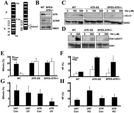
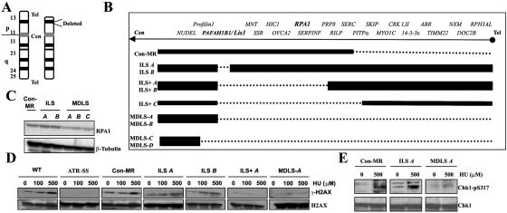
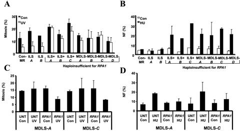
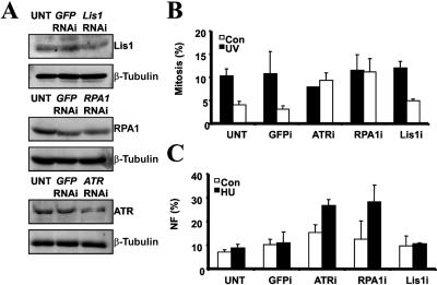
Ataxia telangiectasia and Rad3-related (ATR) protein, a kinase that regulates a DNA damage-response pathway, is mutated in ATR-Seckel syndrome (ATR-SS), a disorder characterized by severe microcephaly and growth delay. Impaired ATR signaling is also observed in cell lines from additional disorders characterized by microcephaly and growth delay, including non-ATR-SS, Nijmegen breakage syndrome, and MCPH1 (microcephaly, primary autosomal recessive, 1)-dependent primary microcephaly. Here, we examined ATR-pathway function in cell lines from three haploinsufficient contiguous gene-deletion disorders--a subset of blepharophimosis-ptosis-epicanthus inversus syndrome, Miller-Dieker lissencephaly syndrome, and Williams-Beuren syndrome--in which the deleted region encompasses ATR, RPA1, and RFC2, respectively. These three genes function in ATR signaling. Cell lines from these disorders displayed an impaired ATR-dependent DNA damage response. Thus, we describe ATR signaling as a pathway unusually sensitive to haploinsufficiency and identify three further human disorders displaying a defective ATR-dependent DNA damage response. The striking correlation of ATR-pathway dysfunction with the presence of microcephaly and growth delay strongly suggests a causal relationship.


Figures

Figure  1.
Figure 1.
BPES-ATR+/− cells, which display an impaired ATR-dependent damage response. A, Chromosome 3 karyotype of the patient with BPES-ATR+/− showing the heterozygous deletion, del(3)(q23,q25). ATR localizes to 3q22-q24. Cen=centromere; Tel=telomere. B, WCE (100 μg) from WT and BPES-ATR+/− LBLs, analyzed by immunoblotting using α-ATR, ATRIP, and NBS1 antibodies. Reduced expression of ATR and ATRIP is seen in BPES-ATR+/− cells specifically. NBS1 served as a loading control and was expressed at normal levels. C, WT, ATR-SS, and BPES-ATR+/− LBLs, exposed to 100 or 500 μM HU for 1 h before chromatin fractionation. ATR-SS and BPES-ATR+/− cells display reduced γ-H2AX compared with WT cells. Blots were reprobed using α-H2AX to confirm loading. D, WT, ATR-SS, and BPES-ATR+/− LBLs, exposed to 500 μM HU for 1 h before extraction. ATR-SS and BPES-ATR+/− cells display reduced Chk1-pS317 formation compared with WT cells. Blots were reprobed using α-Chk1 to confirm loading. E, ATR-SS and BPES-ATR+/− LBLs showing defective UV-induced (5 J/m2) G2/M checkpoint arrest 24 h postirradiation. Arrest in WT LBLs is seen as a decrease in the MI after UV irradiation. F, ATR-SS and BPES-ATR+/− LBLs that, unlike WT cells, show increased NF after 24 h treatment with HU (5 mM). G, UV-induced G2/M defect in BPES-ATR+/− LBLs, complemented after transfection with ATR cDNA. BPES-ATR+/− cells, either untransfected (UNT) or transfected (ATR) with pc-DNA3-ATR, were UV irradiated (5 J/m2), and the MI was determined after 24 h. H, HU-induced NF in BPES-ATR+/−, complemented after transfection with ATR cDNA. BPES-ATR+/− LBLs either untransfected (UNT) or transfected (ATR) with pc-DNA3-ATR were untreated (Con=control) or treated with HU (5 mM) 24 h posttransfection. NF was analyzed 24 h posttreatment with HU.
Figure  2.
Figure 2.
MDLS cells, which display an impaired ATR-dependent DNA damage response. A, Chromosome 17 karyotype of MDLS highlighting the heterozygous deletion at 17p13.3. B, Deletion mapping in the panel of LBLs from patients with ILS, ILS+, and MDLS. The dashed line indicates the heterozygously deleted region. Con-MR is a control LBL from a patient with mild MR who does not exhibit lissencephaly, microcephaly, growth retardation, or MDLS but has a hemizygous telomeric deletion that does not involve either RPA1 or PAFAH1B1/Lis1. ILS A and ILS B denote patients with low-grade ILS due to microdeletions involving PAFAH1B1/Lis1 only. ILS+A, ILS+B, and ILS+C denote patients with larger deletions, a more severe ILS, and additional craniofacial abnormalities, whereas MDLS-A, -B, -C, and -D denote patients with the largest deletions, who exhibit the most severe grade of lissencephaly along with microcephaly and growth retardation. The positions of PAFAH1B1/Lis1 and RPA1 are highlighted. C, Western-blot analysis of RPA1 expression from WCEs from Con-MR, ILS A, ILS B, MDLS-A, MDLS-B, and MDLS-C showing reduced expression of RPA1, specifically in the three MDLS cell lines. D, Defective HU-induced γ-H2AX formation, which segregates with RPA1 haploinsufficiency. Cells were treated as described for figure 1C. Defective γ-H2AX formation is seen in ATR-SS, ILS+ A, and MDLS-A cells, compared with the normal response in WT, Con-MR, ILS A, and ILS B cells. E, Impaired HU-induced Chk1-pS317, seen in MDLSA LBLs, compared with those of Con-MR and ILS A. Cells were treated with 500 μM HU for 1 h before extraction and were reprobed using α-Chk1 to confirm loading.
Figure  3.
Figure 3.
Haploinsufficiency of RPA1, which specifically segregates with a defective ATR-dependent DNA damage response. A, Defective ATR-dependent G2/M checkpoint arrest, which segregates with RPA1 haploinsufficiency. Con-MR, ILS A, and ILS B cells showing a reduction in MI (percentage of mitosis) at 24 h after UV irradiation (5 J/m2), indicating G2/M checkpoint arrest. ILS+ A, ILS+ B, ILS+ C, and MDLS-A, -B, -C, and -D cell lines failed to show a decrease in MI after UV treatment. B, Increased HU-induced NF segregating with RPA1 haploinsufficiency. No increase in HU-induced NF is seen in Con-MR, ILS A, or ILS B cells. In contrast, ILS+ A, ILS+ B, ILS+ C, and MDLS-A, -B, -C, and -D cells show elevated HU-induced NF. C, The ATR-dependent UV-induced G2/M defect in MDLS cells, complemented after transfection with RPA1 cDNA. MDLS-A and MDLS-C cells, either untransfected (“UNT”) or transfected (“RPA1”) with pc-DNA3-RPA1, were unirradiated (Con=control) or UV-irradiated (“UV”) (5 J/m2), and the MI was determined after 24 h. RPA1 cDNA specifically corrected the G2/M checkpoint defect of these cells, as seen by the reduced UV-induced MI after transfection (“RPA1 UV”), compared with untransfected irradiated cells (“UNT UV”). D, The increased HU-induced NF seen in MDLS, complemented after transfection with RPA1 cDNA. MDLS-A and -C, either untransfected (“UNT”), or transfected (“RPA1”) with pc-DNA3-RPA1 were untreated (“Con”) or treated with HU (5 mM) 24 h posttransfection. A reduction in HU-induced NF is specifically seen after transfection of MDLS LBLs with RPA1 cDNA (“RPA1 HU”), compared with the untransfected (“UNT HU”) cells.
Figure  4.
Figure 4.
Inefficient siRNA of ATR or RPA1, which impairs the ATR-dependent DNA damage response. A, WT LBLs transfected once with a low concentration (10 nM) of siRNA oligonucleotides for GFP, Lis1, RPA1, or ATR and analyzed for expression of Lis1, RPA1, and ATR by western blotting, using β-tubulin as a loading control, 24 h posttransfection. B, WT LBLs untransfected (UNT) or transfected with the indicated siRNA oligonucleotides, analyzed for UV-induced G2/M checkpoint arrest by monitoring MI. Transfection with siRNA oligonucleotides against ATR or RPA1 impaired G2/M arrest after UV. In contrast, after transfection with oligonucleotides against GFP and Lis1, an intact G2/M arrest was observed. C, WT LBLs transfected with the indicated siRNA oligonucleotides, analyzed for HU-induced NF. Transfection with siRNA oligonucleotides against ATR and RPA1 caused HU-induced NF, in contrast to the lack of impact of siRNA oligonucleotides against GFP and Lis1.
Figure  5.
Figure 5.
WBS cells, which show an impaired ATR-dependent DNA damage response. A, Chromosome 7 karyotype from WBS showing the location of the submicroscopic heterozygous interstitial deletion on chromosome 7q11.23. B, Deletion mapping of patients with WBS and SVAS showing the position of Elastin (ELN) and RFC2. The dashed line indicates the size of the heterozygous deletion. C, Impaired HU-induced Chk1-pS317, seen in WBS LBLs (WBS-I and WBS-II) compared with those of the clinically normal parent (WT-I and WT-II, respectively). Cells were treated with 500 μM HU for 1 h before extraction and were reprobed using α-Chk1 to confirm loading. D, WBS LBLs, which exhibit impaired UV-induced G2/M checkpoint arrest. The MI was determined 24 h after UV irradiation (5 J/m2). WBS-I and WBS-II LBLs show defective UV-induced G2/M arrest, unlike WT LBLs from their parents (WT-I and WT-II, respectively). E, Increased HU-induced NF, seen in WBS cell lines. LBLs were treated with HU (5 mM) and were examined for NF 24 h after treatment. Both WBS-I and WBS-II LBLs show increased HU-induced NF, unlike WT LBLs from their parents (WT-I and WT-II, respectively). F, UV-induced G2/M defect in WBS LBLs, complemented after transfection with RFC2 cDNA. WBS-I and WBS-II cells, either untransfected (“UNT”) or transfected (“RFC2”) with pc-DNA3-RFC2, were UV irradiated (“UV”) (5 J/m2), and the MI was determined after 24 h. A reduction in MI after UV irradiation is observed after transfection of WBS LBLs with RFC2 cDNA transfection (“RFC2 UV”) compared with the untransfected irradiated cells (“UNT UV”). G, The increased HU-induced NF seen in WBS LBLs, complemented after transfection with RFC2 cDNA. WBS-I and WBS-II, either untransfected (“UNT”) or transfected (“RFC2”) with pc-DNA3-RFC2, were treated with HU (5 mM) 24 h posttransfection. A reduction in HU-induced NF is seen after transfection of WBS LBLs with RFC2 cDNA (“RFC2 HU”), compared with the untransfected HU-treated (“UNT HU”) cells.

References

Web Resource

    1. Online Mendelian Inheritance in Man (OMIM), http://www.ncbi.nlm.nih.gov/Omim/ (for ATR, SS, Nijmegen breakage syndrome, Fanconi anemia, MCPH1, BPES, FOXL2, PAHFAH1B1/Lis1, MDLS, RPA1, WBS, SVAS, ELN, and RFC2)

References

    1. Abraham RT (2001) Cell cycle checkpoint signaling through the ATM and ATR kinases. Genes Dev 15:2177–2196 10.1101/gad.914401 - DOI - PubMed
    1. Shiloh Y (2001) ATM and ATR: networking cellular responses to DNA damage. Curr Opin Genet Dev 11:71–77 10.1016/S0959-437X(00)00159-3 - DOI - PubMed
    1. O’Driscoll M, Ruiz-Perez VL, Woods CG, Jeggo PA, Goodship JA (2003) A splicing mutation affecting expression of ataxia-telangiectasia and Rad3-related protein (ATR) results in Seckel syndrome. Nat Genet 33:497–501 10.1038/ng1129 - DOI - PubMed
    1. O’Driscoll M, Jeggo PA (2003) Clinical impact of ATR checkpoint signalling failure in humans. Cell Cycle 2:194–195 - PubMed
    1. Seckel HPG (1960) Bird-headed dwarfs: studies in developmental anthropology including human proportions. Springer Karger, Basel, Switzerland

Publication types

MeSH terms