Case Reports
. 2007 Jul;81(1):87-103.
doi: 10.1086/519219. Epub 2007 Jun 4.

A new genetic disorder in mitochondrial fatty acid beta-oxidation: ACAD9 deficiency

Affiliations
Case Reports

A new genetic disorder in mitochondrial fatty acid beta-oxidation: ACAD9 deficiency

M He et al. Am J Hum Genet. 2007 Jul.

Abstract

The acyl-CoA dehydrogenases are a family of multimeric flavoenzymes that catalyze the alpha,beta -dehydrogenation of acyl-CoA esters in fatty acid beta -oxidation and amino acid catabolism. Genetic defects have been identified in most of the acyl-CoA dehydrogenases in humans. Acyl-CoA dehydrogenase 9 (ACAD9) is a recently identified acyl-CoA dehydrogenase that demonstrates maximum activity with unsaturated long-chain acyl-CoAs. We now report three cases of ACAD9 deficiency. Patient 1 was a 14-year-old, previously healthy boy who died of a Reye-like episode and cerebellar stroke triggered by a mild viral illness and ingestion of aspirin. Patient 2 was a 10-year-old girl who first presented at age 4 mo with recurrent episodes of acute liver dysfunction and hypoglycemia, with otherwise minor illnesses. Patient 3 was a 4.5-year-old girl who died of cardiomyopathy and whose sibling also died of cardiomyopathy at age 21 mo. Mild chronic neurologic dysfunction was reported in all three patients. Defects in ACAD9 mRNA were identified in the first two patients, and all patients manifested marked defects in ACAD9 protein. Despite a significant overlap of substrate specificity, it appears that ACAD9 and very-long-chain acyl-CoA dehydrogenase are unable to compensate for each other in patients with either deficiency. Studies of the tissue distribution and gene regulation of ACAD9 and very-long-chain acyl-CoA dehydrogenase identify the presence of two independently regulated functional pathways for long-chain fat metabolism, indicating that these two enzymes are likely to be involved in different physiological functions.


Figures
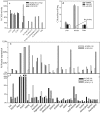
Figure  1.
Figure 1.
Comparison of activity, expression, and tissue localization of VLCAD, ACAD9, and LCAD. The specific activity of VLCAD, ACAD9, and LCAD, as measured with palmitoyl-CoA, was 13, 1.2, and 0.9 U/mg, respectively (a). All substrates used are described in the “Methods” section. b and c, Expression of mRNA of ACAD9 and LCAD (b) and VLCAD, MCAD, and SCAD (c), measured in 21 human tissues by real-time RT-PCR and compared with the relative level of expression in muscle. The ratio of expression of VLCAD, ACAD9, LCAD, MCAD, and SCAD in muscle to the housekeeping gene GUSB was 34, 1.0, 0.4, 4.9, and 17.1, respectively. d, Immunoinactivation of ACAD activity in human liver, muscle, and brain, measured with C18:0-CoA as substrate. VLCAD antibodies inactivated 32%, 100%, and 0% of the activity measured in the mitochondrial membrane fraction of human liver, muscle, and brain, respectively, whereas ACAD9 antibodies inactivated 41%, 0%, and 100% in these same tissues.
Figure  2.
Figure 2.
Immunohistochemical and immunofluorescent staining of normal human brain and muscle sections with ACAD9 antisera and human lung sections with LCAD antisera. a and b, Immunohistochemical staining of human muscle with VLCAD (a) and ACAD9 (b) antibody visualized with DAB (brown), counterstained with a blue nuclear stain. c, Diffuse and punctate pattern of ACAD9 staining, extending to neuronal dendrites of Purkinje cells. The nucleus of Purkinje and granular neurons does not stain with ACAD9 antibodies visualized with fluorescent secondary antibody (white). di, Normal human brain section immunostained with ACAD9 antibody and visualized with DAB (brown). All sections are counterstained with a nuclear stain (blue). d, Neurons of human neocortex. e, Neurons in the CA1 region of normal human hippocampus showing relatively weak ACAD9 staining. f, Neurons in the CA4 region and granular neurons of hippocampus. g, Cerebellar cortex, including Purkinje neurons, their dendrites, and neurons in the granular layer, showing relatively strong ACAD9 staining. h, Absence of ACAD9 staining in cerebellar white matter. i, Strong ACAD9 staining in dentate nucleus in the deep white matter of cerebellum. j and k, Normal human lung sections stained with LCAD (j) or the marker protein MUC1 (k) (a membrane protein, specific to human alveolar septae) antibody and visualized by fluorescent secondary antibodies, LCAD (j, red) and MUC1 ( k, green), colocalizing to type II alveolar epithelial cells (white arrow), as described in the “Methods” section. l and m, LCAD antibodies specifically staining bronchial epithelium and the alveolar septae of normal human lung tissue (l). At high magnification, the staining of LCAD in bronchial epithelium (m) shows a diffused punctate pattern consistent with mitochondrial staining.
Figure  3.
Figure 3.
Immunoinactivation of purified ACAD9 protein. Serial dilutions of purified rabbit anti-ACAD9 IgG were used to inactivate 1.5 μg of purified recombinant ACAD9 or VLCAD. Reactions were performed in the presence of 0.5 mg/ml of BSA and 5% glycerol. After incubation with antibody, reactions were precipitated with S. aureus protein A–conjugated Sepharose beads, and the remaining ACAD activity was measured with the ETF fluorescence-reduction assay. The data plotted represent the average of duplicate measurements. a, Twenty to 80 μg of ACAD9 antibody incubated with purified ACAD9. The Y-axis shows the relative activity compared with enzyme without antibody treatment. b, Up to 160 μg of ACAD9 antibody incubated with purified VLCAD protein and plotted as was that in panel a.
Figure  4.
Figure 4.
Promoter function of the ACAD9 gene and its putative regulatory pathway. a, Sequence of the human ACAD9 gene promoter region, as predicted by computational analysis. The transcriptional start sites (TSS1 and TSS2) were inferred from the consensus of the 5′ end of EST tags in dbEST in combination with PCR analysis. b, Two conserved nucleotide blocks that match the consensus sequences for NRF-1 and CRE transcription-factor binding sites. The dotted line indicates the invariant nucleotides in the recognition sites among different species. The NRF-1 and CRE sites are duplicated in all primates. c, A series of fragments containing increasing lengths of 5′ UTR from the ACAD9 gene, inserted upstream of a firefly luciferase reporter gene (Luc) in the promoterless pGL3-basic vector. The TAAG insertion identified in the patient is shown as a vertical bar in exon 1. d, The promoter-luciferase fusion constructs, transfected into HepG2 cells along with an internal control. The luciferase activity is presented as the mean and SD from at least three independent experiments, as described in the “Methods” section. The 4-bp insertion was introduced in plasmid p3A9 (e), and the activities were measured as were those in panel d.
Figure  5.
Figure 5.
Subcellular location of ACAD9 protein. a and b, Western blot of fibroblast and liver lysate with anti-human ACAD9 antibodies. One major band (M) in control samples (a, lanes 2 and 4; b, lane 2) co-migrated with purified recombinant mitochondrial ACAD9 (a, lane 1; b, lane 1), and another was larger (c/n). Both forms were missing or reduced in patient samples (a, lanes 3 and 5; b, lane 3). VLCAD-deficient fibroblasts (V) (a, lane 2) and normal human fibroblast (N) (b, lane 2) or postmortem liver (N) (a, lane 4) served as controls. ce, Immunofluorescent staining of normal fibroblasts with ACAD9 (c, green) and mitochondrial ATP synthesase (mATPase) (d, red) antibodies, visualized by fluorescent secondary antibodies. The merged image (e) shows that the two proteins colocalize in part (yellow). A green ACAD9 signal is seen in the nucleus (white arrow). Panels f–h and i–k are the same as panels c–e but use fibroblasts from patients 2 and 1 (P2 and P1), respectively. ln, Immunofluorescent staining of postmortem liver from patient 3 (P3) and a control (C) with antibodies against ACAD9 (l, green) or liver-specific α1-AT (m, red), visualized by fluorescent secondary antibodies. The merged signal is shown in panel n. ACAD9 (αA9) (lanes 1 and 2) staining is deficient in patient 3 (lane 2), but MCAD (αM) (lane 3) and VLCAD (αV) (lane 4) staining are normal. ot, Immunofluorescent staining of postmortem cerebellar cortex of patient 1 (P1) (oq) and control (rt) with antibodies to the cerebellar cortex marker EAAT4 (o and r, red) or ACAD9 (p and s, green); the merged signal is shown in panels q and t, in pseudocolored yellow. ACAD9 is prominent in control (s) but is absent in patient cerebellum (p). A white arrow indicates ACAD9-specific staining of dentate nucleus in deep white matter of the normal control.
Figure  6.
Figure 6.
Immunocompetition and subcellular fractionation of ACAD9 antigen in human fibroblast lysate. a, Immunocompetition of ACAD9 antigen in human fibroblast lysate by purified ACAD9 protein. Fifty micrograms of normal human fibroblast lysate or 40 ng of purified ACAD9 protein were separated by SDS-PAGE and were transferred to a PVDF membrane. The membrane was then incubated with ACAD9 antibodies (affinity purified) with and without an equal amount (mole/mole) of purified ACAD9 protein. Lanes 1 and 2 show the reaction of fibroblast extract and purified ACAD9 with ACAD9 antibody. Lanes 3 and 4 show the same as do lanes 1 and 2, except that the ACAD9 signals on the membrane are diminished when purified ACAD9 protein is included in the antibody-incubation mixture. Arrows point to two ACAD9 protein species with different molecular masses. The band marked “mA9” corresponds in size to purified mature mitochondrial ACAD9, whereas the other ACAD9 species (c/n A9) is ∼10 kDa bigger than mA9 and is of a size consistent with the predicted cytosolic/nuclear form of ACAD9. Lane 5, Molecular mass markers. The two marked bands are β-galactosidase (94 kDa) and albumin (66 kDa). b, Subcellular distribution of ACAD9 proteins in normal fibroblast extract. Fibroblasts were lysed by repeated cycles of freezing and thawing, and the cytoplasmic fraction (lane 4) was separated from the nuclear/mitochondrial fraction (lane 3) by centrifugation. Lane 2, Purified ACAD9. Protein markers in lane 1 are the same as those in panel a.
Figure  7.
Figure 7.
Molecular analysis of ACAD9-deficient patients. a, ACAD9 consisting of 22 exons and generating two predominant molecular species through alternative splicing. The schematic drawing illustrates that the basic ACAD domain structure of both translated proteins is conserved (ACDN, ACDM, and ACDC); however, in one, a nuclear targeting signal (N) is substituted for the mitochondrial targeting signal (M). This variant message also contains a longer exon 1, giving a longer N-terminus (U) and a shorter exon 18 along with additional exons 19–22. b and c, Amplification of ACAD9 sequences from control (C), patient 1 (P1), and patient 2 (P2) mRNA. PCRs with control fibroblast cDNA as template readily amplified full-length ACAD9 coding sequences (b, lane 6; c, lane 2 ) and overlapping subfragments (b, lane 2, exons 1–7; b, lane 4, exons 6–18). Full-length message (exons 1–18) could not be amplified from mRNA either from patient 1 (b, lane 7) or patient 2 (c, lane 3). Shorter-than-expected fragments were amplified from patient 1 cDNA (b, lane 3, exons 1–7; lane 5, exons 6–18). d, Sequence of ACAD9 UTR from genomic DNA and cDNA. Sequencing chromatograms of genomic DNA from patient 1 (left panel) identified a dual pattern due to a 4-bp insertion. Patient cDNA in that region showed only the normal sequence, without the insertion (right panel). e, Diagrams of ACAD9 species amplified from cDNA from patient 1. One consisted of exon 1 joined to exon 6 (α), whereas the other consisted of exon 11 jointed to exon 18 (β).
Figure  8.
Figure 8.
Mild perivascular chronic inflammatory cell cuffing in postmortem patient brain. Scattered perivascular lymphocytes (arrows) cuff the blood vessels in the brain stem of patient 1. a, Hematoxylin and eosin stain (640× magnification) and the neocortical white matter. b, LCA immunohistochemistry stain (640× magnification). This inflammatory cell infiltrate is also accompanied by some hemosiderin-laden macrophages, seen only in the deep white matter of the centrum semiovale and the brain stem but not in gray matter or leptomeninges where the acute damage was most severe. Neither of these findings appears to be related to the child’s acute demise; rather, they appear to be chronic. These changes are reminiscent of, though not as severe as, those reported in X-linked adrenoleukodystrophy (MIM 300100).

References

Web Resources

    1. AceView, http://www.ncbi.nlm.nih.gov/IEB/Research/Acembly/
    1. BLAST, http://www.ncbi.nlm.nih.gov/blast/
    1. dbEST, http://www.ncbi.nlm.nih.gov/dbEST/
    1. dbSNP, http://www.ncbi.nlm.nih.gov/SNP/
    1. GenBank, http://www.ncbi.nlm.nih.gov/Genbank/ (for full-length human ACAD9 cDNA sequence from mRNA variant b [accession number NM_014049] and partial human ACAD9 cDNA sequence from mRNA variant a [accession numbers CR613592 and BX415793])

References

    1. Vockley J, Whiteman DA (2002) Defects of mitochondrial β-oxidation: a growing group of disorders. Neuromuscul Disord 12:235–24610.1016/S0960-8966(01)00308-X - DOI - PubMed
    1. Spiekerkoetter U, Sun B, Zytkovicz T, Wanders R, Strauss AW, Wendel U (2003) MS/MS-based newborn and family screening detects asymptomatic patients with very-long-chain acyl-CoA dehydrogenase deficiency. J Pediatr 143:335–34210.1067/S0022-3476(03)00292-0 - DOI - PubMed
    1. Furuta S, Miyazawa S, Hashimoto T (1981) Purification and properties of rat liver acyl-CoA dehydrogenases and electron transfer flavoprotein. J Biochem (Tokyo) 90:1739–1750 - PubMed
    1. Ensenauer R, He M, Willard JM, Goetzman ES, Corydon TJ, Vandahl BB, Mohsen AW, Isaya G, Vockley J (2005) Human acyl-CoA dehydrogenase-9 plays a novel role in the mitochondrial beta-oxidation of unsaturated fatty acids. J Biol Chem 280:32309–3231610.1074/jbc.M504460200 - DOI - PubMed
    1. Zhang J, Zhang W, Zou D, Chen G, Wan T, Zhang M, Cao X (2002) Cloning and functional characterization of ACAD-9, a novel member of human acyl-CoA dehydrogenase family. Biochem Biophys Res Commun 297:1033–104210.1016/S0006-291X(02)02336-7 - DOI - PubMed

Publication types

MeSH terms

Associated data