. 2007 Jul;81(1):136-46.
doi: 10.1086/518902. Epub 2007 May 14.

The novel neuronal ceroid lipofuscinosis gene MFSD8 encodes a putative lysosomal transporter

Affiliations

The novel neuronal ceroid lipofuscinosis gene MFSD8 encodes a putative lysosomal transporter

Eija Siintola et al. Am J Hum Genet. 2007 Jul.

Abstract

The late-infantile-onset forms are the most genetically heterogeneous group among the autosomal recessively inherited neurodegenerative disorders, the neuronal ceroid lipofuscinoses (NCLs). The Turkish variant was initially considered to be a distinct genetic entity, with clinical presentation similar to that of other forms of late-infantile-onset NCL (LINCL), including age at onset from 2 to 7 years, epileptic seizures, psychomotor deterioration, myoclonus, loss of vision, and premature death. However, Turkish variant LINCL was recently found to be genetically heterogeneous, because mutations in two genes, CLN6 and CLN8, were identified to underlie the disease phenotype in a subset of patients. After a genomewide scan with single-nucleotide-polymorphism markers and homozygosity mapping in nine Turkish families and one Indian family, not linked to any of the known NCL loci, we mapped a novel variant LINCL locus to chromosome 4q28.1-q28.2 in five families. We identified six different mutations in the MFSD8 gene (previously denoted "MGC33302"), which encodes a novel polytopic 518-amino acid membrane protein that belongs to the major facilitator superfamily of transporter proteins. MFSD8 is expressed ubiquitously, with several alternatively spliced variants. Like the majority of the previously identified NCL proteins, MFSD8 localizes mainly to the lysosomal compartment. However, the function of MFSD8 remains to be elucidated. Analysis of the genome-scan data suggests the existence of at least three more genes in the remaining five families, further corroborating the great genetic heterogeneity of LINCLs.


Figures
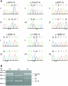
Figure  1.
Figure 1.
MFSD8-associated mutations. A, Sequence chromatograms showing homozygous MFSD8 mutations in six patients with vLINCL (f3, e3, j3, c3, h3, and b3) and the respective control (c) sequences. The mutated bases are underlined, and the vertical lines indicate exon-intron boundaries. B, Aberrant splicing of the c.754+2T→A mutant transcript. The agarose gel shows the mRNA splicing pattern in patient e5 (A/A), who is homozygous for c.754+2T→A, in his heterozygous carrier mother (T/A), and in a control individual (T/T), analyzed by RT-PCR with primers from exons 6 and 11. In the sample from patient e5, an altered pattern of PCR products is seen, with almost complete lack of the normal transcript (∼550 bp) containing exons 7–10, a complete lack of an alternatively spliced variant lacking exon 7 (Δ7) (∼400 bp), and increased expression of two alternatively spliced variants: one lacking exon 8 (Δ8) (∼500 bp) and one lacking exons 7 and 8 (Δ7+8) (∼350 bp). In addition, a faint band of ∼480 bp, the sequence of which remains unknown, is visible in the samples from patient e5 and his mother. The positions of the 100-bp ladder-size marker are shown on the left.
Figure  2.
Figure 2.
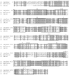
Conservation of the MFSD8 amino acid sequence among human (Homo sapiens), mouse (Mus musculus), and zebrafish (Danio rerio). Amino acid sequences were aligned using MAFFT version 5.8. The amino acid numbering is indicated for the human polypeptide. Identical amino acids are indicated with gray shading. Sites of exonic disease-causing mutations are marked with an asterisk (*): p.Arg233Gly/splice, p.Tyr298X, p.Gly310Asp, p.Asp368His/splice, and p.Gly429Asp. The predicted 12 transmembrane domains are indicated with boxes and Roman numerals.
Figure  3.
Figure 3.
Northern-blot analysis of MFSD8 expression. An ∼5-kb MFSD8 transcript (upper panels) was detected in all tissues analyzed. In all brain regions and in lung, it is the only transcript seen. In other tissues, transcripts that ranged in size from ∼1 to ∼3 kb and/or ∼6 kb were also present. Lower panels, Hybridization with human β-actin cDNA as a control for RNA loading. The positions of size markers are shown on the left of each blot.
Figure  4.
Figure 4.
Schematic structure of the MFSD8 protein, with positions of patient mutations that affect exonic sequences. MFSD8 is predicted to contain 12 transmembrane domains, according to the prediction programs HMMTOP, TMHMM, and TMpred. The site of the nonsense mutation (p.Tyr298X) is indicated with a square, the site of the missense mutations (p.Gly310Asp and p.Gly429Asp) with circles, and the site of mutations that either change amino acids or affect splicing (p.Arg233Gly/splice and p.Asp368His/splice) with crosses.
Figure  5.
Figure 5.
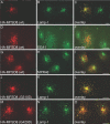
Lysosomal localization of both wild-type and mutant MFSD8 proteins. Subcellular localization of HA-tagged wild-type (wt) and mutant (G310D and G429D) MFSD8 was seen in transiently transfected COS-1 cells. A, D, and G, Distribution of the HA-MFSD8 protein by use of an antibody against HA (red). B, Lysosomal staining pattern by use of an anti–Lamp-1 antibody (green). C, Overlay of the HA-MFSD8 and Lamp-1 stainings. E, Early endosomal staining pattern by use of an anti-EEA1 antibody (green). F, Overlay of the HA-MFSD8 and EEA1 stainings. H, Trans-Golgi-network staining pattern by use of an anti-MPR46 antibody (green). I, Overlay of the HA-MFSD8 and MPR46 stainings. J and M, Distribution of the G310D mutant (J) and the G429D mutant (M) HA-MFSD8 proteins by use of an antibody against HA (red). K and N, Lysosomal staining pattern by use of an anti–Lamp-1 antibody (green). L and O, Overlays of the mutant HA-MFSD8 and Lamp-1 stainings. Yellow indicates overlap of wild-type or mutant HA-MFSD8 and the subcellular markers. Scale bars (10 μm) are shown in panels C, F, I, L, and O.

References

Web Resources

    1. BLAST, http://www.ncbi.nlm.nih.gov/BLAST/ (for NCBI protein-protein BLAST)
    1. FlyBase, http://flybase.bio.indiana.edu/
    1. GenBank, http://www.ncbi.nlm.nih.gov/Genbank/ (for MFSD8 [accession number NM_152778])
    1. HMMTOP, http://www.enzim.hu/hmmtop/
    1. MAFFT version 5.8, http://align.bmr.kyushu-u.ac.jp/mafft/online/server/

References

    1. Santavuori P (1988) Neuronal ceroid-lipofuscinoses in childhood. Brain Dev 10:80–83 - PubMed
    1. Haltia M (2003) The neuronal ceroid-lipofuscinoses. J Neuropathol Exp Neurol 62:1–13 - PubMed
    1. The International Batten Disease Consortium (1995) Isolation of a novel gene underlying Batten disease, CLN3. Cell 82:949–95710.1016/0092-8674(95)90274-0 - DOI - PubMed
    1. Vesa J, Hellsten E, Verkruyse LA, Camp LA, Rapola J, Santavuori P, Hofmann SL, Peltonen L (1995) Mutations in the palmitoyl protein thioesterase gene causing infantile neuronal ceroid lipofuscinosis. Nature 376:584–58710.1038/376584a0 - DOI - PubMed
    1. Sleat DE, Donnelly RJ, Lackland H, Liu CG, Sohar I, Pullarkat RK, Lobel P (1997) Association of mutations in a lysosomal protein with classical late-infantile neuronal ceroid lipofuscinosis. Science 277:1802–180510.1126/science.277.5333.1802 - DOI - PubMed

Publication types

MeSH terms

Associated data