. 2007 Jul;81(1):147-57.
doi: 10.1086/518426. Epub 2007 May 24.

Recurrent mutation in the first zinc finger of the orphan nuclear receptor NR2E3 causes autosomal dominant retinitis pigmentosa

Affiliations

Recurrent mutation in the first zinc finger of the orphan nuclear receptor NR2E3 causes autosomal dominant retinitis pigmentosa

Frauke Coppieters et al. Am J Hum Genet. 2007 Jul.

Abstract

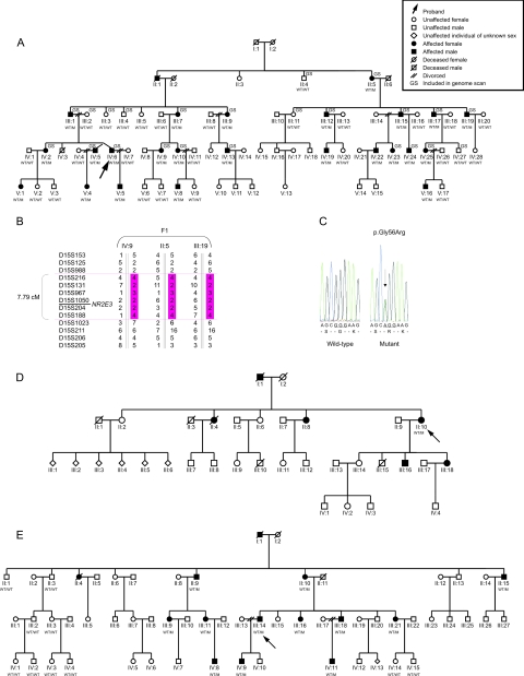
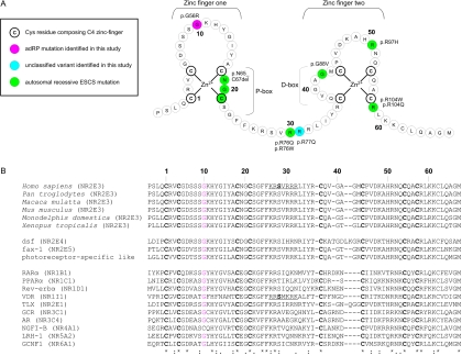
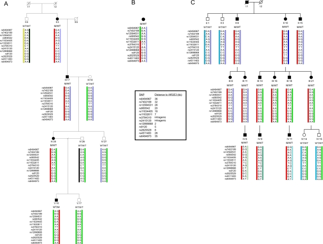
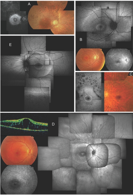
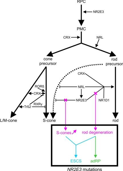
"Autosomal dominant retinitis pigmentosa" (adRP) refers to a genetically heterogeneous group of retinal dystrophies, in which 54% of all cases can be attributed to 17 disease loci. Here, we describe the localization and identification of the photoreceptor cell-specific nuclear receptor gene NR2E3 as a novel disease locus and gene for adRP. A heterozygous mutation c.166G-->A (p.Gly56Arg) was identified in the first zinc finger of NR2E3 in a large Belgian family affected with adRP. Overall, this missense mutation was found in 3 families affected with adRP among 87 unrelated families with potentially dominant retinal dystrophies (3.4%), of which 47 were affected with RP (6.4%). Interestingly, affected members of these families display a novel recognizable NR2E3-related clinical subtype of adRP. Other mutations of NR2E3 have previously been shown to cause autosomal recessive enhanced S-cone syndrome, a specific retinal phenotype. We propose a different pathogenetic mechanism for these distinct dominant and recessive phenotypes, which may be attributed to the dual key role of NR2E3 in the regulation of photoreceptor-specific genes during rod development and maintenance.


Figures

Figure  1.
Figure  1.
A, Pedigree of F1, in which NR2E3 mutation p.Gly56Arg was found. WT = wild type; M = mutation p.Gly56Arg. B, Haplotypes of critical recombinants of F1 used to narrow candidate region. C, Sequence electropherogram of heterozygous substitution c.166G→A (p.Gly56Arg) versus wild type. D and E, Pedigrees of F2 and F3, respectively.
Figure  2.
Figure  2.
A, Theoretical model of the two C4 zinc fingers located in the DBD of NR2E3. The first zinc finger contains the P-box, in which two or three exposed residues are responsible for half-site sequence recognition. The second zinc finger harbors a dimerization interface for DBDs (D-box) and is involved in recognition of half-site spacing and mutual orientation. A consensus numbering system, starting from the first Cys of the first zinc finger, is used for comparison with other NRs in panel B. B, top rows, Alignment of the human protein sequence of NR2E3 with five orthologs. Middle rows, Three nonhuman NRs belonging to the same subfamily as NR2E3 (dsf, D. melanogaster; fax-1, C. elegans; and photoreceptor-specific like, D. melanogaster). Bottom rows, Human NRs belonging to one of six NR subfamilies: (1) Thyroid hormone receptor–like (RARα, PPARα, Rev-erbα and VDR); (2) HNF4-like (TLX); (3) Estrogen receptor–like (GR and AR); (4) Nerve Growth factor IB receptor–like (NGFI-B); (5) Fushi tarazu-F1–like (LRH-1); and (6) Germ cell nuclear factor–like (GCNF1). The official NR nomenclature is given in parentheses. Cys residues composing the C4 zinc-finger motif are indicated in bold. Gly residues in other NRs, corresponding to Gly56 in NR2E3, are indicated in red. Gly56 is conserved in almost all members of the human NR superfamily and in different orthologs, suggesting a functional constraint. Underlined sequences indicate a nuclear localization signal (NLS) in VDR and the corresponding putative NLS in NR2E3. The amino acid Ser, in bold, represents Ser74 and Ser51 in NR2E3 and VDR, respectively (details provided in table 3).
Figure  3.
Figure  3.
Haplotype analysis with intragenic and closely flanking SNPs. Genotyping in a core family of F1 (A), the proband of F2 (B), and a core family of F3 (C), performed using 2 intragenic SNPs and 11 flanking SNPs that are located in a region of 70 kb around the 7.7-kb NR2E3 gene (HapMap, Ensembl). In F1 and F3, the red bar represents the common disease haplotype. In F2, this disease haplotype is partially shared from SNP rs11632611 (located 7 kb upstream of NR2E3) to SNP rs6494973 (35 kb downstream of NR2E3), as represented by the red bar. The green bar represents the distinct haplotype block (ranging from SNP rs11634405 at 18 kb to SNP rs6494967 at 36 kb both upstream of NR2E3). Genotyping of an anonymous microsatellite at 50 kb downstream of NR2E3 (UCSC Genome Browser, Primer3) allowed delineation of a maximal common region of ∼75 kb (upstream rs11634405 at 18 kb – NR2E3 7.7 kb – downstream microsatellite at 50 kb) (data not shown). WT, wild type; M, mutant. Filled bars represent affected individuals. Pedigree numbers are represented below the symbols and are in agreement with the numbering of figure 1.
Figure  4.
Figure  4.
Full-field flash ERG. A, ERG traces of patient IV:24 of F1 at age 13 years. Scotopic (dark-adapted) rod-specific responses are completely absent, whereas scotopic maximal combined rod-cone responses are derived only from cones; amplitudes of photopic (light-adapted) transient and 30-Hz flicker cone-specific responses are moderately reduced, with limited latency delays; aspects of scotopic maximal combined rod-cone responses and photopic cone-specific responses to a transient flash are not identical. B, ERG traces of patient III:15 of F1 at age 62 years. All responses are absent, illustrating that gross retinal function is unmeasurable. C, Normal traces, for comparison. All ERGs are according to standards of International Society for Clinical Electrophysiology of Vision (ISCEV). RE, right eye; LE, left eye; μV/div, microvolts per division.
Figure  5.
Figure  5.
Clinical characteristics of affected members of F1 and F3, revealing a novel NR2E3-related clinical subtype of adRP. A, Late-stage phenotype in patient III:15 of F1 at age 62 years; composite autofluorescence (AF) (left) and composite fundus (right) of right eye (RE). Note small, relatively well-preserved macular area on fundus picture—although bull’s-eye pattern of hyperautofluorescence can be seen on AF in that area, with two hyperfluorescent rings that have virtually merged into one; also note severe narrowing of retinal vasculature. B, Early-stage phenotype in patient IV:24 of F1 at age 13 years; composite of AF (top) and fundus (bottom left) and late-phase fluorescein angiography (FA) of posterior pole of RE. Note the presence of two hyperautofluorescent rings on AF: one in bull’s-eye pattern around the fovea, another in midperiphery beyond temporal vascular arcades and nasal to optic disc (OD). Also note diffuse chorioretinal atrophy in areas corresponding to hyperautofluorescent rings visible on fundus and angiography picture. The small darker spot above the superior temporal vascular arcade on AF corresponds to a small retinal hemorrhage unrelated to RP phenotype. Discrete narrowing of retinal vasculature is also shown. C, Midstage phenotype in patient IV:23 of F1 at age 31 years. Composite AF (left) and composite fundus pictures (right) of posterior pole of left eye (LE) shows a bull’s-eye pattern with one small, discrete, intensely hyperautofluorescent ring around the fovea and a second, more diffusely hyperautofluorescent ring within area of temporal vascular arcades. Also note nummular areas of hypoautofluorescence above temporal vascular arcade, corresponding to areas of outer retinal atrophy; a dark string-like opacity superotemporal between vascular arcades which corresponds to a floater; marked attenuation of retinal vasculature; choroidal vasculature shining through due to diffuse chorioretinal atrophy, without intraretinal pigmentation; and a good-quality fovea. D, Midstage phenotype in patient IV:9 of F3. Clockwise from right, composite infrared image (IR) of fundus of RE, late phase FA and fundus picture of posterior pole, and optical coherence tomography (OCT) of central macular area of RE at age 16 years. Note cystic degeneration of fovea on IR, fundus picture, and OCT, without progressive fluorescein diffusion (leakage), unlike cystic macular edema, on FA, but identical to cystic degeneration seen in ESCS. IR further shows two whiter concentric rings in bull’s-eye pattern around the fovea, with the larger ring located just inside the temporal vascular arcades; also note sparse spicular pigmentation in inferior periphery and mild attenuation of retinal vasculature. E, Midstage phenotype in patient IV:19 of F1 at age 29 years; composite AF of RE. Note three concentric rings around posterior pole: one in a bull’s-eye pattern around fovea, a second at level of temporal vascular arcades and nasal to OD, and a third in periphery (only depicted inferiorly); hypoautofluorescence due to outer retinal degeneration beyond the peripheral hyperautofluorescent ring; and moderate vascular attenuation.
Figure  6.
Figure  6.
Role of retinal TFs during photoreceptor development. Several TFs are required to determine a specific photoreceptor cell type (rod or cone). First, multipotent retinal progenitor cells (RPC) pass through different stages of competence, finally becoming postmitotic cells (PMC). The expression of CRX commits a population of PMCs toward photoreceptor lineage. A pool of these are directed to cone fate. It is presumed that cone precursors follow an S-cone default pathway (influenced by CRX and RORβ18,19) and that subsequent L- and M-cone induction is mediated by additional TFs. One of these is Trβ2, inducing M-opsin expression but repressing S-opsin expression, probably as a heterodimer with the nuclear receptor RXRγ., Later in development, another pool of PMCs will acquire the rod fate by NRL expression. NRL activates transcription of NR2E3, having a positive synergistic influence with CRX, NR1D1, and NRL on the transcription of rod-specific genes., Moreover, NR2E3 contributes to rodlike morphology. Importantly, rod precursors are relatively “plastic,” because they are still capable of developing S-cones. NRL and especially NR2E3 are able to repress transcription of cone-specific genes, to stabilize the rod fate before further development of rodlike characteristics.,, This dual function of NR2E3, involving both transrepression of cone-specific genes and transactivation of rod-specific genes, may underlie molecular pathogenesis of distinct NR2E3-related retinal dystrophies, as illustrated in this study. We hypothesize that mutations affecting both functions lead to an increased density of S-cones and a degeneration of rods, as seen in ESCS, whereas mutations affecting only or predominantly the transactivation function result in a predominant degeneration of rods, as seen in adRP. Recent studies have suggested that temporal differences exist between those NR2E3 functions. NR2E3 expression has been shown in mitotic as well as postmitotic cells of the developing retina., Moreover, NR2E3 expression was also seen in both rods and cones of the mature retina. It is presumed that in postmitotic rods NR2E3 influences rod cell differentiation, whereas, in late mitotic progenitors, its primary function is to suppress the cone-generation program.

References

Web Resources

    1. Berkeley Drosophila Genome Project (BDGP), http://www.fruitfly.org/seq_tools/splice.html
    1. Ensembl Genome Browser, http://www.ensembl.org/index.html
    1. ESEfinder, http://rulai.cshl.edu/tools/ESE/
    1. International HapMap Project, http://www.hapmap.org/
    1. NCBI Map Viewer, http://www.ncbi.nlm.nih.gov/mapview/static/MVstart.html

References

    1. Bird AC (1995) Retinal photoreceptor dystrophies LI. Am J Ophthalmol 119:543–562 - PubMed
    1. Sullivan LS, Bowne SJ, Birch DG, Hughbanks-Wheaton D, Heckenlively JR, Lewis RA, Garcia CA, Ruiz RS, Blanton SH, Northrup H, et al (2006) Prevalence of disease-causing mutations in families with autosomal dominant retinitis pigmentosa: a screen of known genes in 200 families. Invest Ophthalmol Vis Sci 47:3052–306410.1167/iovs.05-1443 - DOI - PMC - PubMed
    1. Lindner TH, Hoffmann K (2005) easyLINKAGE: a PERL script for easy and automated two-/multi-point linkage analyses. Bioinformatics 21:405–40710.1093/bioinformatics/bti009 - DOI - PubMed
    1. Mykytyn K, Braun T, Carmi R, Haider NB, Searby CC, Shastri M, Beck G, Wright AF, Iannaccone A, Elbedour K, et al (2001) Identification of the gene that, when mutated, causes the human obesity syndrome BBS4. Nat Genet 28:188–19110.1038/88925 - DOI - PubMed
    1. Haider NB, Jacobson SG, Cideciyan AV, Swiderski R, Streb LM, Searby C, Beck G, Hockey R, Hanna DB, Gorman S, et al (2000) Mutation of a nuclear receptor gene, NR2E3, causes enhanced S cone syndrome, a disorder of retinal cell fate. Nat Genet 24:127–13110.1038/72777 - DOI - PubMed

Substances