. 2007 Jul;81(1):170-9.
doi: 10.1086/519494. Epub 2007 Jun 4.

Pleiotropic effects of CEP290 (NPHP6) mutations extend to Meckel syndrome

Affiliations

Pleiotropic effects of CEP290 (NPHP6) mutations extend to Meckel syndrome

Lekbir Baala et al. Am J Hum Genet. 2007 Jul.

Abstract

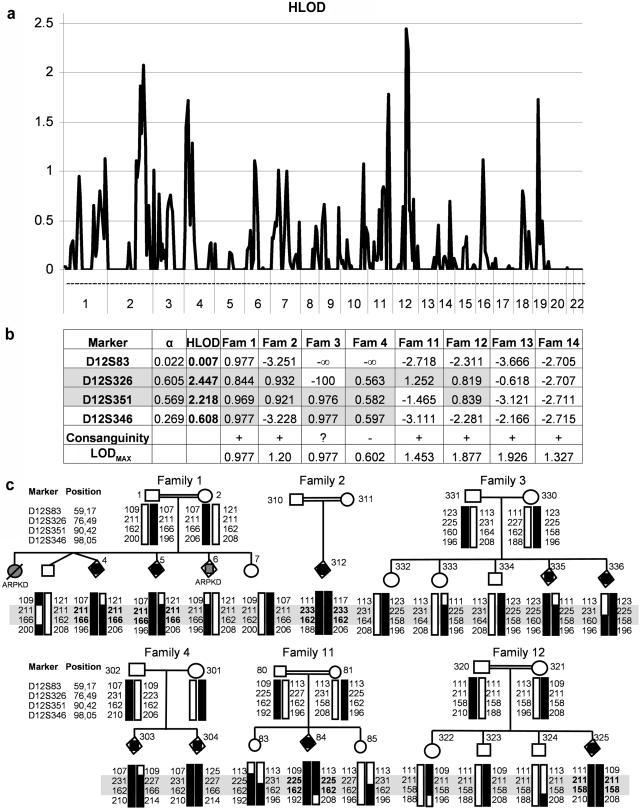
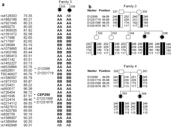
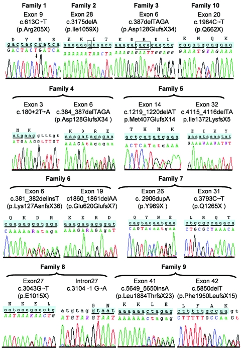
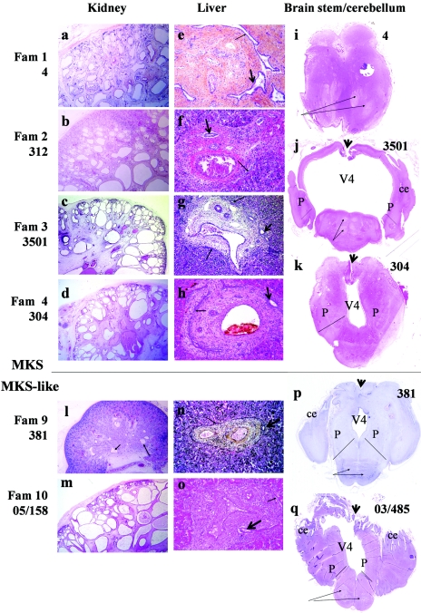
Meckel syndrome (MKS) is a rare autosomal recessive lethal condition characterized by central nervous system malformations, polydactyly, multicystic kidney dysplasia, and ductal changes of the liver. Three loci have been mapped (MKS1-MKS3), and two genes have been identified (MKS1/FLJ20345 and MKS3/TMEM67), whereas the gene at the MKS2 locus remains unknown. To identify new MKS loci, a genomewide linkage scan was performed using 10-cM-resolution microsatellite markers in eight families. The highest heterogeneity LOD score was obtained for chromosome 12, in an interval containing CEP290, a gene recently identified as causative of Joubert syndrome (JS) and isolated Leber congenital amaurosis. In view of our recent findings of allelism, at the MKS3 locus, between these two disorders, CEP290 was considered a candidate, and homozygous or compound heterozygous truncating mutations were identified in four families. Sequencing of additional cases identified CEP290 mutations in two fetuses with MKS and in four families presenting a cerebro-reno-digital syndrome, with a phenotype overlapping MKS and JS, further demonstrating that MKS and JS can be variable expressions of the same ciliopathy. These data identify a fourth locus for MKS (MKS4) and the CEP290 gene as responsible for MKS.


Figures

Figure  1.
Figure 1.
Genomewide scan. a, Results of the multipoint linkage analysis with microsatellite markers performed in eight families (Fam), with use of MERLIN software under the assumption of a fully penetrant recessive model with a disease-allele of frequency of 0.0001 and with allowance for heterogeneity between families. The highest HLOD score (2.45) was found at marker D12S326. b, Summary of HLOD observed at chromosome 12 in each of the eight families. Six of the eight families show a LOD score at marker D12S326, D12S351, or D12S346 close to its maximal value (LODmax). c, Haplotyping at the 12q21 locus in the six families showing potential linkage to chromosome 12. Homozygosity for two consecutive markers—D12S326 and D12S351 (boxed)—is observed in five affected siblings of four families (1, 2, 11, and 12). In family 1, the first child and case 6 (gray) are affected with ARPKD.
Figure  2.
Figure 2.
Haplotype analysis of families 3 and 4. a, Haplotyping analysis with Affymetrix 10K SNP chips of affected siblings 335 and 336 of family 3, which identified a single homozygous region on chromosome 12 between markers rs1882186 and rs1389064. This 10-Mb region includes CEP290. b, Analysis of four microsatellite markers flanking CEP290 in family 3, which confirmed homozygosity in only affected siblings 335 and 336 and reduced the telomeric boundary of the interval to position 88.71 (D12S1678). In family 4 carrying the same maternally inherited p.Asp128GlufsX34 mutation as family 3, the maternal disease haplotype is different from fetuses 335 and 336 and does not suggest a founder effect for the mutation. Positions of SNP and microsatellite markers are given according to the UCSC Genome Browser database.
Figure  3.
Figure 3.
Sequence chromatographs of CEP290 mutations identified in the present study. Mutation numbering is based on cDNA sequence, where +1 corresponds to the A of the ATG translation codon in the GenBank cDNA reference sequence NM_025114.3. The predicted effect of the mutation on the protein is also given with the amino acid number.
Figure  4.
Figure 4.
Pathological features of fetal cases with CEP290 mutations. Histological sections (HES staining) of kidney (left), liver (middle), and brain stem/cerebellum (right) of MKS- and MKS-like–affected fetuses with CEP290 mutations. In families 1–4 (a–d) and 10 (m), kidney histology shows major abnormalities characteristic of MKS: cysts in both kidney and medulla, growing from periphery to center. Small areas of conserved nephrogenesis are observed in the subcapsular zone. In family 9 (l), kidney histology shows conserved corticomedullary organization with several generations of mature glomeruli. Microcysts are found in the deep cortex, but tubular microcysts are observed mainly in medulla (black arrows). Liver histology shows a portal fibrosis with persistent ductal plate (arrows) and/or BDP/dilatation (arrowheads). Subject 4 (b) had the most-severe liver fibroadenomatosis, whereas the liver anomalies were moderate and focal in families 9 (n) and 10 (o). Brain-stem/cerebellum transverse sections are shown for five cases. i, Subject 4: section of the mesencephalon showing the chaotic organization of fibers and tracts (arrows) and atretic aqueduct of Sylvius. j, Subject 3501: section at the level of the pons showing severely dilated V4, enlarged cerebellar peduncle (P), chaotic organization of fibers and tracts (arrows), hypoplastic vermis (arrowhead), and cerebellar hemispheres (ce). k, Fetus 304. Note the inverted MTS with dysmorphic V4 flanked with enlarged and elongated cerebellar peduncles (P). For fetuses 381 (p) and 03/485 (q), the section at the level of the pons shows an inverted MTS with chaotic fibers and tracts (arrows), dysmorphic V4, severe hypoplasia of vermis (arrowhead), and well-developed cerebellar hemispheres. Fam=family.

References

Web Resources

    1. GenBank, http://ncbi.nlm.nih.gov/Genbank/ (for cDNA translation codon [accession number NM_025114.3])
    1. Online Mendelian Inheritance in Man (OMIM), http://www.ncbi.nlm.nih.gov/Omim/ (for MKS, Meckel-like/Goldston syndrome, JS, LCA, and BBS)
    1. UCSC Genome Browser, http://genome.ucsc.edu/

References

    1. Kyttala M, Tallila J, Salonen R, Kopra O, Kohlschmidt N, Paavola-Sakki P, Peltonen L, Kestila M (2006) MKS1, encoding a component of the flagellar apparatus basal body proteome, is mutated in Meckel syndrome. Nat Genet 38:155–15710.1038/ng1714 - DOI - PubMed
    1. Smith UM, Consugar M, Tee LJ, McKee BM, Maina EN, Whelan S, Morgan NV, Goranson E, Gissen P, Lilliquist S, et al (2006) The transmembrane protein meckelin (MKS3) is mutated in Meckel-Gruber syndrome and the wpk rat. Nat Genet 38:191–19610.1038/ng1713 - DOI - PubMed
    1. Dawe HR, Smith UM, Cullinane AR, Gerrelli D, Cox P, Badano JL, Blair-Reid S, Sriram N, Katsanis N, Attie-Bitach T, et al (2007) The Meckel-Gruber syndrome proteins MKS1 and meckelin interact and are required for primary cilium formation. Hum Mol Genet 16:173–18610.1093/hmg/ddl459 - DOI - PubMed
    1. Roume J, Genin E, Cormier-Daire V, Ma HW, Mehaye B, Attie T, Razavi-Encha F, Fallet-Bianco C, Buenerd A, Clerget-Darpoux F, et al (1998) A gene for Meckel syndrome maps to chromosome 11q13. Am J Hum Genet 63:1095–1101 - PMC - PubMed
    1. Khaddour R, Smith U, Baala L, Martinovic J, Clavering D, Shaffiq R, Ozilou C, Cullinane A, Kyttälä M, Shalev S, et al (2007) Spectrum of MKS1 and MKS3 mutations in Meckel syndrome: a genotype-phenotype correlation. Hum Mutat 28:523–52410.1002/humu.9489 - DOI - PubMed

Publication types

MeSH terms

Associated data