

CT visible internal stone structure, but not Hounsfield unit value, of calcium oxalate monohydrate (COM) calculi predicts lithotripsy fragility in vitro
- PMID: 17565491
- PMCID: PMC2408919
- DOI: 10.1007/s00240-007-0104-6
CT visible internal stone structure, but not Hounsfield unit value, of calcium oxalate monohydrate (COM) calculi predicts lithotripsy fragility in vitro
Abstract
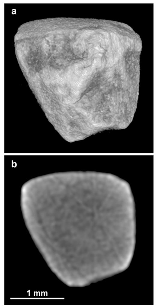
Calcium oxalate monohydrate (COM) stones are often resistant to breakage using shock wave (SW) lithotripsy. It would be useful to identify by computed tomography (CT) those COM stones that are susceptible to SW's. For this study, 47 COM stones (4-10 mm in diameter) were scanned with micro CT to verify composition and also for assessment of heterogeneity (presence of pronounced lobulation, voids, or apatite inclusions) by blinded observers. Stones were then placed in water and scanned using 64-channel helical CT. As with micro CT, heterogeneity was assessed by blinded observers, using high-bone viewing windows. Then stones were broken in a lithotripter (Dornier Doli-50) over 2 mm mesh, and SW's counted. Results showed that classification of stones using micro CT was highly repeatable among observers (kappa = 0.81), and also predictive of stone fragility. Stones graded as homogeneous required 1,874 +/- 821 SW/g for comminution, while stones with visible structure required half as many SW/g, 912 +/- 678. Similarly, when stones were graded by appearance on helical CT, classification was repeatable (kappa = 0.40), and homogeneous stones required more SW's for comminution than did heterogeneous stones (1,702 +/- 993 SW/g, compared to 907 +/- 773). Stone fragility normalized to stone size did not correlate with Hounsfield units (P = 0.85). In conclusion, COM stones of homogeneous structure require almost twice as many SW's to comminute than stones of similar mineral composition that exhibit internal structural features that are visible by CT. This suggests that stone fragility in patients could be predicted using pre-treatment CT imaging. The findings also show that Hounsfield unit values of COM stones did not correlate with stone fragility. Thus, it is stone morphology, rather than X-ray attenuation, which correlates with fragility to SW's in this common stone type.
Figures


References
-
- Schubert G. Stone analysis. Urol Res. 2006;34:146–150. - PubMed
-
- Daudon M, Donsimoni R, Hennequin C, Fellahi S, Le Moel G, Paris M, Troupel S, Lacour B. Sex and age-related composition of 10617 calculi analyzed by infrared-spectroscopy. Urol Res. 1995;23:319–326. - PubMed
-
- Bon D, Dore B, Irani J, Marroncle M, Aubert J. Radiographic prognostic criteria for extracorporeal shock-wave lithotripsy: a study of 485 patients. Urology. 1996;48:556–560. discussion 560-551. - PubMed
-
- Dretler SP. Stone fragility—a new therapeutic distinction. J Urol. 1988;139:1124–1127. - PubMed
-
- Williams JC, Jr., Saw KC, Paterson RF, Hatt EK, McAteer JA, Lingeman JE. Variability of renal stone fragility in shock wave lithotripsy. Urology. 2003;61:1092–1096. - PubMed
Publication types
MeSH terms
Substances
Grants and funding
LinkOut - more resources
Full Text Sources
Other Literature Sources
Research Materials
