

Experiences and benefits of positron emitted tomography-computed tomography (PET-CT) combined with video-assisted thoracoscopic surgery (VATS) in the diagnosis of Stage 1 sarcoidosis
- PMID: 17565511
- PMCID: PMC1879162
- DOI: 10.1631/jzus.2007.B0410
Experiences and benefits of positron emitted tomography-computed tomography (PET-CT) combined with video-assisted thoracoscopic surgery (VATS) in the diagnosis of Stage 1 sarcoidosis
Abstract
Background: The purpose of this study was to describe our experiences and analyze the benefits of video-assisted thoracoscopic surgery (VATS) combined with positron emitted tomography (PET)-computed tomography (CT) in the diagnosis of patients with early (Stage 1) sarcoidosis.
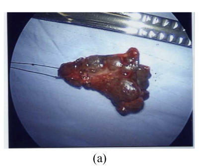
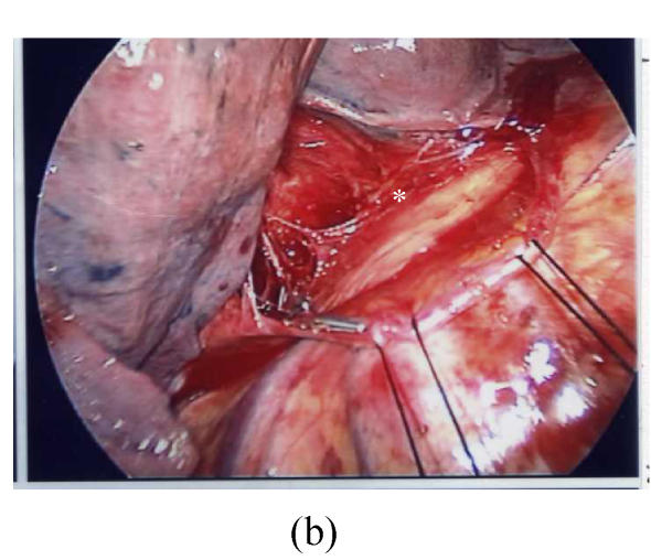
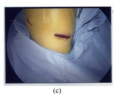
Methods: From 1995 to 2006, seven patients (two males, five females), with ages ranging from 26 to 58 years, were impressed with Stage 1 sarcoidosis (mediastinal or hilar lymph nodes involvements without lung involvement) by histological examination of intrathoracic lymph nodes (LNs) and/or lung parenchyma taken from VATS biopsy. Three of them received PET or PET-CT evaluation. VATS was approached from the right and left side in one and six patients, respectively, according to the locations of their lesions.
Results: All the VATS biopsied LNs or lung specimens were adequate for establishing diagnosis. Mediastinal LNs were taken from Groups 3, 4 in four, Group 7 in two, and Groups 5, 6 in one of them. Hilar LNs biopsies were performed in four cases. Lung biopsy was performed in all but two cases. All of them were expressed pathologically or radiologically as Stage 1 sarcoidosis. PET-CT revealed high emission signals over these affected LNs. These patients received oral steroid treatment or follow up only. All of them were followed up from 5 months to 11 years with satisfactory results.
Conclusion: VATS biopsy is a minimally invasive, safe and effective procedure. It can be used as a diagnostic alternative of transbronchial lung biopsy (TBLB), and can harvest larger and more areas of specimens than mediastinoscopy for staging patients with sarcoidosis. PET-CT can provide us more accurate information about the characteristics and localization of these lesions before biopsy. VATS combined with PET-CT can provide more accurate and earlier diagnosis of patients with unknown intrathoracic lesions, including the sarcoidosis.
Figures


Similar articles
-
Preoperative intrathoracic lymph node staging in patients with non-small-cell lung cancer: accuracy of integrated positron emission tomography and computed tomography.Eur J Cardiothorac Surg. 2009 Sep;36(3):440-5. doi: 10.1016/j.ejcts.2009.04.003. Epub 2009 May 22. Eur J Cardiothorac Surg. 2009. PMID: 19464906 Review.
-
Transbronchial needle aspiration accurately diagnoses subcentimetre mediastinal and hilar lymph nodes detected by integrated positron emission tomography and computed tomography.Respirology. 2007 Nov;12(6):848-55. doi: 10.1111/j.1440-1843.2007.01164.x. Respirology. 2007. PMID: 17986113
-
[Indication of video-assisted thoracic surgery for mediastinal mass lesions].Kyobu Geka. 2012 Oct;65(11):934-8. Kyobu Geka. 2012. PMID: 23023535 Japanese.
-
Accuracy of 18F-FDG PET/CT for lymph node staging in non-small-cell lung cancers.Chin Med J (Engl). 2009 Aug 5;122(15):1749-54. Chin Med J (Engl). 2009. PMID: 19781319
-
[A case of sarcoidosis presenting as a solitary pulmonary nodule].Nihon Kokyuki Gakkai Zasshi. 2008 Dec;46(12):992-6. Nihon Kokyuki Gakkai Zasshi. 2008. PMID: 19195199 Review. Japanese.
Cited by
-
In Which Patients with Sarcoidosis Is FDG PET/CT Indicated?J Clin Med. 2020 Mar 24;9(3):890. doi: 10.3390/jcm9030890. J Clin Med. 2020. PMID: 32213991 Free PMC article. Review.
-
Sarcoidosis and its otolaryngological implications.Eur Arch Otorhinolaryngol. 2010 Oct;267(10):1507-14. doi: 10.1007/s00405-010-1331-y. Epub 2010 Jul 9. Eur Arch Otorhinolaryngol. 2010. PMID: 20617327 Review.
-
Video-assisted thoracoscopic surgery (VATS) for the treatment of hepatic hydrothorax: report of twelve cases.J Zhejiang Univ Sci B. 2009 Jul;10(7):547-51. doi: 10.1631/jzus.B0820374. J Zhejiang Univ Sci B. 2009. PMID: 19585673 Free PMC article.
-
Video-assisted thoracoscopic surgery (VATS) for bilateral primary spontaneous pneumothorax.J Zhejiang Univ Sci B. 2008 Apr;9(4):335-40. doi: 10.1631/jzus.B0720235. J Zhejiang Univ Sci B. 2008. PMID: 18381810 Free PMC article.
References
-
- ATS (American Thoracic Society) Statement on Sarcoidosis. Joint Statement of the American Thoracic Society (ATS), the European Respiratory Society (ERS) and the World Association of Sarcoidosis and Other Granulomatous Disorders (WASOG) adopted by the ATS Board of Directors and by the ERS Executive Committee. February 1999. Am J Respir Crit Care Med. 1999;160(2):736–755. - PubMed
-
- Beyer T, Townsend DW, Brun T, Kinahan PE, Charron M, Roddy R, Jerin J, Young J, Byars L, Nutt R. A combined PET/CT scanner for clinical oncology. J Nucl Med. 2000;41:1369–1379. - PubMed
-
- Brigid GA, Flanagan FL, Dehdashti F. Whole-body positron emission tomography: normal variations, pitfalls, and technical considerations. AJR. 1997;169:1675–1680. - PubMed
-
- Cosabel U. Sarcoidosis: clinical update. Eur Respir J Suppl. 2001;18(32):56s–68s. - PubMed
-
- Geraint James D, Figueroa Lebron RE. Update on sarcoidosis. Bol Asoc Med P R. 1979;71:325–335. - PubMed
