. 2007 Jun 12:8:22.
doi: 10.1186/1471-2121-8-22.

Glycosylation, transport, and complex formation of palmitoyl protein thioesterase 1 (PPT1)--distinct characteristics in neurons

Affiliations

Glycosylation, transport, and complex formation of palmitoyl protein thioesterase 1 (PPT1)--distinct characteristics in neurons

Annina Lyly et al. BMC Cell Biol. .

Abstract

Background: Neuronal ceroid lipofuscinoses (NCLs) are collectively the most common type of recessively inherited childhood encephalopathies. The most severe form of NCL, infantile neuronal ceroid lipofuscinosis (INCL), is caused by mutations in the CLN1 gene, resulting in a deficiency of the lysosomal enzyme, palmitoyl protein thioesterase 1 (PPT1). The deficiency of PPT1 causes a specific death of neocortical neurons by a mechanism, which is currently unclear. To understand the function of PPT1 in more detail, we have further analyzed the basic properties of the protein, especially focusing on possible differences in non-neuronal and neuronal cells.

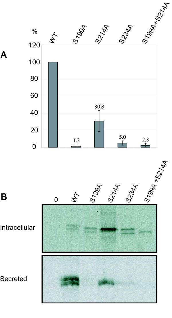
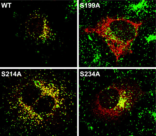
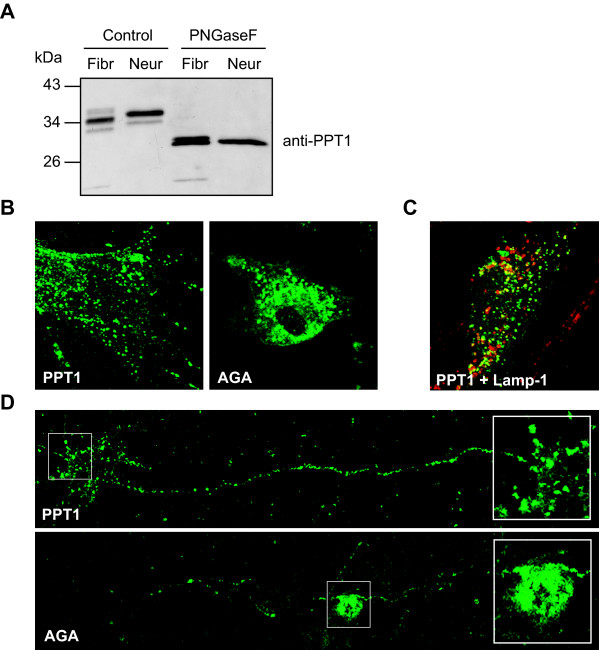
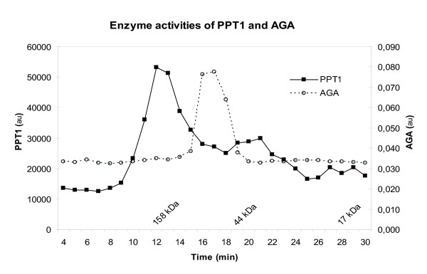
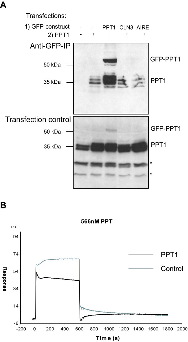
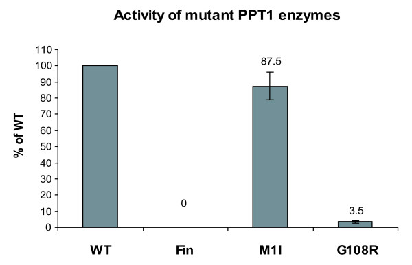
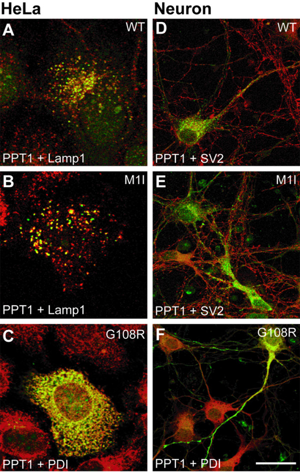
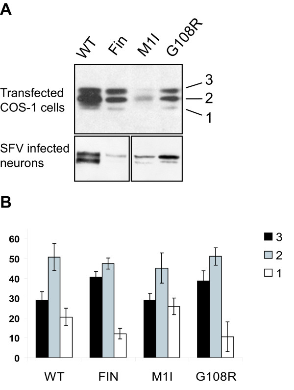
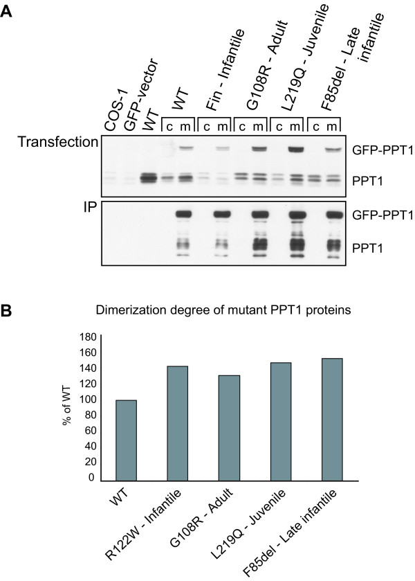
Results: Our study shows that the N-glycosylation of N197 and N232, but not N212, is essential for PPT1's activity and intracellular transport. Deglycosylation of overexpressed PPT1 produced in neurons and fibroblasts demonstrates differentially modified PPT1 in different cell types. Furthermore, antibody internalization assays showed differences in PPT1 transport when compared with a thoroughly characterized lysosomal enzyme aspartylglucosaminidase (AGA), an important observation potentially influencing therapeutic strategies. PPT1 was also demonstrated to form oligomers by size-exclusion chromatography and co-immunoprecipitation assays. Finally, the consequences of disease mutations were analyzed in the perspective of our new results, suggesting that the mutations increase both the degree of glycosylation of PPT1 and its ability to form complexes.

Conclusion: Our current study describes novel properties for PPT1. We observe differences in PPT1 processing and trafficking in neuronal and non-neuronal cells, and describe for the first time the ability of PPT1 to form complexes. Understanding the basic characteristics of PPT1 is fundamental in order to clarify the molecular pathogenesis behind neurodegeneration in INCL.


Figures

Figure 1
Figure 1
Activity and processing of wild type and glycosylation site mutated PPT1. (A) COS-1 cells were transfected with plasmids producing mutated PPT1 enzymes and lysed 48 h post-transfection. PPT1 enzyme activity of each lysate (5 μg total protein) was analyzed by a one-hour reaction in + 37°C. Transfection efficacies were controlled by immunofluorescence analysis. The data shown represents an average of three independent experiments and the standard deviations between these experiments are marked as error bars. (B) COS-1 cells were transfected and metabolically labelled with 35S-Cysteine for 1 hour, chased for 2 hours, immunoprecipitated, separated in SDS-PAGE and autoradiographed. Both intracellular and secreted forms of PPT are shown.
Figure 2
Figure 2
Intracellular localization of wild type and glycosylation site mutated PPT1 in HeLa cells. HeLa cells were transfected with the wild type pCMV5-PPT1 plasmid or with plasmids carrying mutations S199A, S214A, S214A and S199A + S214A. The cells were double-stained using the polyclonal antibody for PPT1 (red) and the monoclonal antibody for LAMP-1, lysosomal membrane protein (green). Colocalization is shown in yellow.
Figure 3
Figure 3
Adenovirus-mediated PPT1 in fibroblasts and neurons. (A) Adenovirus-mediated PPT1 was expressed in mouse fibroblasts and neurons. Cell lysates (5 μg of total protein) were treated with PNGaseF to remove the N-glycans from the polypeptides. Proteins were separated with SDS-PAGE and analyzed by immunoblotting. Antibody internalization assay was performed in adenovirus infected mouse fibroblasts (B) and neurons (D) with both AGA and PPT1 (green). Double staining with the lysosomal marker LAMP-1 (red) is shown for internalized PPT1 in fibroblasts (C). Co-localization is shown in yellow.
Figure 4
Figure 4
Size-exclusion chromatography of fractionated PC12 cells: enzyme activities of PPT1 and AGA. PC12 cells were homogenized and the 10 000g pellet of the postnuclear supernatant was isolated by centrifugation. Solubilised fraction was subjected to size exclusion chromatography and the PPT1 and AGA enzyme activities were measured from the fractions collected. The X-axis indicates time and the molecular weights are derived from the standard (Bio-Rad).
Figure 5
Figure 5
Oligomerization of PPT1. (A) Cells were transfected with wild type pCMV5-PPT1 plasmids or/and with plasmids producing a GFP-fusion protein as indicated (GFP-PPT1, GFP-CLN3 or GFP-AIRE). The lysates were immunoprecipitated with anti-GFP-conjugated agarose beads. Both lysates (Transfection control) and immunoprecipitated samples (Anti-GFP-IP) were analyzed by immunoblotting using the anti-PPT1 antibody. * = Unspecific binding. (B) Biacore assay investigating the self-interaction of purified PPT1 (see methods).
Figure 6
Figure 6
Enzyme activity of PPT1 with disease mutations in transfected COS-1 cells. COS-1 cells were transfected with plasmids producing mutated PPT1 enzymes. The cells were lysed 48 h post-transfection and the enzyme activity of each lysate (5 μg of total protein) was analyzed by a one-hour reaction in + 37°C. The data was normalized for PPT1 expression by immunoblotting and densitometric scanning. Here the background activity is assumed to be zero. The data shown represents an average of three independent experiments and the standard deviations between these experiments are marked as error bars.
Figure 7
Figure 7
Intracellular localization of wild type and mutant PPT1 in non-neuronal and neuronal cells. HeLa cells (A-C) transfected with pCMV5-PPT1 plasmids and neurons (D-F) infected with PPT1-SFV bearing the indicated mutations were double-stained for PPT1 using the GST-PPT1 antibody (green), for lysosomes using the Lamp-1 antibody (red), for synaptic vesicles using the SV2 antibody (red) and for ER using the PDI antibody (red). Colocalization is shown in yellow. Scale bar = 20 μm.
Figure 8
Figure 8
Glycosylation degree of mutant PPT1 molecules. (A) 10 μg of total protein from the cell lysates of transfected COS-1 cells and SFV infected neurons was analyzed by immunoblotting and densitometric scanning. The numbers 3, 2 and 1 indicate the three differently glycosylated forms of PPT1. (B) The graph represents relative amounts of tri-, di-, and monoglycosylated forms of PPT1 in transfected COS-1 cells. The data shown represents an average of three independent experiments and the error bars show the standard deviations between these experiments.
Figure 9
Figure 9
Oligomerization rate of wild type and mutant PPT1 molecules. (A) COS-1 cells were either single or double transfected with pCMV5-hPPT1 or with plasmids having the indicated mutations, together with GFP-vector (c) or with the corresponding GFP-PPT1 construct having the same mutation in the PPT molecule (m). The cells were lysed 48 h post-transfection. To verify the transfections, 10 μg of total protein from the lysates was analyzed by SDS-PAGE and immunoblotting using both anti-GFP antibody (not shown) and anti-PPT1 antibody (Transfection). The lysates were then subjected to immunoprecipitation using anti-GFP-agarose beads. Immunoprecipitates were analyzed by SDS-PAGE and immunoblotting using the anti-PPT1 antibody (IP). (B) The graph represents the dimerization degree of mutant proteins compared to wild type (calculated as a ratio between the immunoprecipitated GFP-PPT/PPT and the transfected GFP-PPT/PPT). An example of a single experiment's quantification is shown experiments.

Similar articles

Cited by

References

    1. Santavuori P, Haltia M, Rapola J, Raitta C. Infantile type of so-called neuronal ceroid-lipofuscinosis. 1. A clinical study of 15 patients. J Neurol Sci. 1973;18:257–267. doi: 10.1016/0022-510X(73)90075-0. - DOI - PubMed
    1. Haltia M, Rapola J, Santavuori P. Infantile type of so-called neuronal ceroid-lipofuscinosis. Histological and electron microscopic studies. Acta Neuropathol (Berl) 1973;26:157–170. doi: 10.1007/BF00697751. - DOI - PubMed
    1. Vesa J, Hellsten E, Verkruyse LA, Camp LA, Rapola J, Santavuori P, Hofmann SL, Peltonen L. Mutations in the palmitoyl protein thioesterase gene causing infantile neuronal ceroid lipofuscinosis. Nature. 1995;376:584–587. doi: 10.1038/376584a0. - DOI - PubMed
    1. Verkruyse LA, Hofmann SL. Lysosomal targeting of palmitoyl-protein thioesterase. J Biol Chem. 1996;271:15831–15836. doi: 10.1074/jbc.271.26.15831. - DOI - PubMed
    1. Hellsten E, Vesa J, Olkkonen VM, Jalanko A, Peltonen L. Human palmitoyl protein thioesterase: evidence for lysosomal targeting of the enzyme and disturbed cellular routing in infantile neuronal ceroid lipofuscinosis. Embo J. 1996;15:5240–5245. - PMC - PubMed

Publication types

MeSH terms

LinkOut - more resources