. 2007 Jun 19;104(25):10482-7.
doi: 10.1073/pnas.0704360104. Epub 2007 Jun 12.

Reversine increases the plasticity of lineage-committed mammalian cells

Affiliations

Reversine increases the plasticity of lineage-committed mammalian cells

Shuibing Chen et al. Proc Natl Acad Sci U S A. .

Abstract

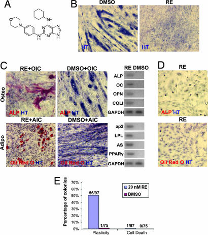
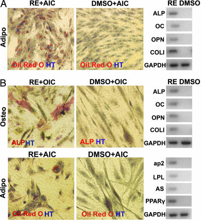
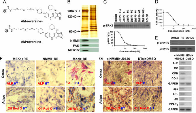
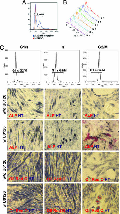
Previously, a small molecule, reversine, was identified that reverses lineage-committed murine myoblasts to a more primitive multipotent state. Here, we show that reversine can increase the plasticity of C2C12 myoblasts at the single-cell level and that reversine-treated cells gain the ability to differentiate into osteoblasts and adipocytes under lineage-specific inducing conditions. Moreover, reversine is active in multiple cell types, including 3T3E1 osteoblasts and human primary skeletal myoblasts. Biochemical and cellular experiments suggest that reversine functions as a dual inhibitor of nonmuscle myosin II heavy chain and MEK1, and that both activities are required for reversine's effect. Inhibition of MEK1 and nonmuscle myosin II heavy chain results in altered cell cycle and changes in histone acetylation status, but other factors also may contribute to the activity of reversine, including activation of the PI3K signaling pathway.


Conflict of interest statement

The authors declare no conflict of interest.

Figures

Fig. 1.
Fig. 1.
Reversine (RE) increases the cellular plasticity of C2C12 myoblasts. (A) Chemical structure of reversine. (B) Reversine blocks the terminal differentiation of C2C12 myoblasts. C2C12 myoblasts (plated at 6,000 cells per cm2) were cultured in GM in the presence or absence of 20 nM reversine for 4 days and then stained with hematoxylin (HT). (C) C2C12 myoblasts gain multipotency after 48-h treatment with 20 nM reversine. C2C12 myoblasts (plated at 6,000 cells per cm2) were cultured in GM supplemented with 20 nM reversine for 48 h. After removal of compound, cells were cultured in OI or AI conditions (OIC or AIC) for an additional 6 days and analyzed by histocytochemistry and RT-PCR. (D) Reversine does not induce C2C12 myoblasts to directly transdifferentiate into osteoblasts or adipocytes without LSICs. After 48-h treatment with 20 nM reversine, cells were fixed and analyzed by histocytochemistry. (E) Clonal analysis. Reversine increases the cellular plasticity of C2C12 myoblasts at the single-cell level. C2C12 myoblasts (plated at 1 cell per well in 96-well plate with GM) were grown in the presence of 20 nM reversine for 2 weeks. The resulting colonies were split into two and cultured with OI or AI conditions for an additional 6 days. Plasticity, cells with increased plasticity that can differentiate into adipocytes and osteoblasts; Cell Death, the colonies that underwent apoptosis during the expanding process. DMSO was used as a negative control in all experiments. Red, ALP, Oil red O; blue, HT.
Fig. 2.
Fig. 2.
Reversine increases cellular plasticity of 3T3E1 osteoblasts and hSMs. (A) The 3T3E1 osteoblasts (plated at 6,000 cells per cm2 in GM) were cultured in the presence of 300 nM reversine (RE) for 4 days. The compound was then removed, and cells were grown in AI conditions for an additional 6 days. (B) hSMs cells (plated at 3,000 cells per cm2 in GM) were cultured in the presence of 300 nM reversine for 4 days. The compound was then removed, and cells were grown in OI or AI conditions (OIC or AIC) for an additional 9 days. The cells were analyzed by histocytochemistry and RT-PCR. DMSO was used as a negative control in all experiments. Red, ALP, Oil red O; blue, HT.
Fig. 3.
Fig. 3.
Affinity chromatography and target identification. (A) Structures of positive (AM·reversine+) and negative (AM·reversine−) affinity matrices. (B) Silver staining and Western blot analysis of proteins retained by affinity supports. (C and D) Reversine inhibits the enzymatic activity of active MEK1 in a dose-dependent fashion as indicated by ERK2-phosphorylation (C) and reversine inhibits the ATPase activity of myosin II (heavy chain) in vitro (D). (E) Reversine (20 nM) blocks MEK-dependent ERK1/2-phosphorylation in C2C12 cells. C2C12 cells were treated with 20 nM reversine for 1 h, and cells were lysed and analyzed by Western blot. DMSO was used as a negative control. U0126 (10 μM) was used as a positive control. (F) Overexpression of MEK1 or NMMII can partially block reversine's activity. C2C12 myoblasts (plated at 6,000 cells per cm2 in GM) transiently transfected with MEK1 or NMMII (with FuGENE6) were cultured in the presence of 20 nM reversine for 48 h. The compound was then removed, and cells were cultured in OI or AI conditions for an additional 6 days and analyzed by histocytochemistry. Empty vector was used as a negative control (Mock). (G) C2C12 myoblasts transfected with siNMMIIs (with X-treme) and cultured in the presence of 10 μM U0126 in GM for 48 h gain multipotency. C2C12 myoblasts (plated at 6,000 cells per cm2 in GM) transiently transfected with siNMMIIs were cultured in the presence of 10 μM U0126 for 48 h. The compound was then removed, and the cells were cultured in OI or AI conditions for an additional 6 days and analyzed by histocytochemistry. Cells transfected with NTsi and treated with DMSO were used as a negative control. Red, ALP, Oil red O; blue, HT.
Fig. 4.
Fig. 4.
Cell cycle effects. (A) Treatment with reversine induces G2/M phase accumulation in C2C12 myoblasts. C2C12 myoblasts treated with 20 nM reversine in GM for 48 h were analyzed by FACS based on DNA content (by using PI staining). (B) C2C12 myoblasts were cultured in GM in the presence of 20 nM reversine for 48 h. After removal of reversine (0 h), cells cultured in GM were analyzed by FACS. (C) C2C12 myoblasts synchronized at G2/M phase and treated with 10 μM U0126 gain multipotency. C2C12 myoblasts synchronized at G1/S, S, or G2/M phase were analyzed by FACS. After release from synchronization by the addition of fresh GM in the presence or absence of 10 μM U0126, cells were incubated for 4 h. The compound was then removed, and the cells were cultured in OI or AI conditions for an additional 6 days and analyzed by histocytochemistry. Red, ALP, Oil red O; blue, HT.

References

    1. Hochedlinger K, Jaenisch R. Nature. 2006;441:1061–1067. - PubMed
    1. Wilmut I, Schnieke AE, McWhir J, Kind AJ, Campbell KH. Nature. 1997;385:810–813. - PubMed
    1. Cowan CA, Atienza J, Melton DA, Eggan K. Science. 2005;309:1369–1373. - PubMed
    1. Tada M, Takahama Y, Abe K, Nakatsuji N, Tada T. Curr Biol. 2001;11:1553–1558. - PubMed
    1. Takahashi K, Yamanaka S. Cell. 2006;126:663–676. - PubMed

Publication types

LinkOut - more resources