. 2007 Jul 10;46(27):8024-35.
doi: 10.1021/bi700341y. Epub 2007 Jun 13.

Probing the role of the histidine 759 ligand in cobalamin-dependent methionine synthase

Affiliations

Probing the role of the histidine 759 ligand in cobalamin-dependent methionine synthase

Matthew D Liptak et al. Biochemistry. .

Abstract

Cobalamin-dependent methionine synthase (MetH) of Escherichia coli is a 136 kDa, modular enzyme that undergoes large conformational changes as it uses a cobalamin cofactor as a donor or acceptor in three separate methyl transfer reactions. At different points during the reaction cycle, the coordination to the cobalt of the cobalamin changes; most notably, the imidazole side chain of His759 that coordinates to the cobalamin in the "His-on" state can dissociate to produce a "His-off" state. Here, two distinct species of the cob(II)alamin-bound His759Gly variant have been identified and separated. Limited proteolysis with trypsin was employed to demonstrate that the two species differ in protein conformation. Magnetic circular dichroism and electron paramagnetic resonance spectroscopies were used to show that the two species also differ with respect to the axial coordination to the central cobalt ion of the cobalamin cofactor. One form appears to be in a conformation poised for reductive methylation with adenosylmethionine; this form was readily reduced to cob(I)alamin and subsequently methylated [albeit yielding a unique, five-coordinate methylcob(III)alamin species]. Our spectroscopic data revealed that this form contains a five-coordinate cob(II)alamin species, with a water molecule as an axial ligand to the cobalt. The other form appears to be in a catalytic conformation and could not be reduced to cob(I)alamin under any of the conditions tested, which precluded conversion to the methylcob(III)alamin state. This form was found to possess an effectively four-coordinate cob(II)alamin species that has neither water nor histidine coordinated to the cobalt center. The formation of this four-coordinate cob(II)alamin "dead-end" species in the His759Gly variant illustrates the importance of the His759 residue in governing the equilibria between the different conformations of MetH.


Figures
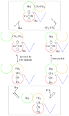
Figure 1
Figure 1
Schematic representation of the catalytic cycle of MetH. The four modules of MetH are colored green (Hcy-binding), gold (CH3-H4folate-binding), red (Cbl-binding), and blue (AdoMet-binding; used for reactivation). The Hcy- and CH3-H4folate-binding modules are rigidly fixed with respect to one another and only one of these two modules can interact with the Cbl at any given time, requiring large conformational changes to provide the Cbl access to the two different substrates during turnover. Substrate binding and product release can only occur on the module that is not interacting with the Cbl-binding module; the dotted boxes show the conformation formed after substrate binding and prior the conformational change required for product release. Methyl transfer from MeCbl to Hcy to form Met and Co1+Cbl is irreversible (6), while transfer from MeCbl to tetrahydrofolate (FH4) is reversible.
Figure 2
Figure 2
Schematic representation of the catalytic and reactivation cycles of MetH. The scheme depicting catalytic turnover (lower left) has been simplified by omitting the substrate binding and product release steps, steps contained in the boxes in Figure 1. As indicated in this figure by the dashed arrow, the Co1+Cbl cofactor becomes oxidized about once in every 2000 turnovers (26), yielding a Co2+Cbl species that is in the His-on form. Addition of Fld results in a switch to His-off Co2+Cbl and conversion from a catalytic conformation (dotted box on the left) to the reactivation conformation (dotted box on the right). Following electron transfer from reduced Fld and methyl transfer from AdoMet, His-off MeCbl is formed. The subsequent conversion to His-on MeCbl with MetH in a catalytic conformation is the rate-limiting step in the reductive methylation reaction (11). As indicated by the broken arrows, enzyme in the Co1+Cbl-bound form is unable to interconvert between catalytic and reactivation conformations (13).
Figure 3
Figure 3
FPLC trace of Co2+Cbl-bound H759G MetH, obtained using a MonoQ 16/10 column. The two fractions labeled Peak 1 and Peak 2 correspond to H759G(cat) and H759G(act), respectively.
Figure 4
Figure 4
SDS-PAGE gels of the products obtained by partial proteolysis of the two distinct FPLC fractions of Co2+Cbl-bound H759G MetH. In each case the enzyme was cleaved into three major fragments: the Cbl-binding module (28 kDa, red), AdoMet-binding module (38 kDa, blue), and combined Hcy- and CH3-H4folate-binding modules (70 kDa, green and yellow). However, while in the case of fraction 1 (top panel) the fragments corresponding to the Cbl-binding module and the combined Hcy- and CH3-H4folate-binding modules eluted together, the Cbl- and AdoMet-binding modules eluted together when fraction 2 was used (bottom panel).
Figure 5
Figure 5
4.5 K Abs spectra of: (a) aqueous Co2+Cbl, (b) aqueous Co2+Cbi+, (c) Co2+Cbl-bound H759G MetH(act), and (d) Co2+Cbl-bound H759G MetH(cat).
Figure 6
Figure 6
7T, 4.5 K MCD spectra of: (a) aqueous Co2+Cbl, (b) aqueous Co2+Cbi+, (c) Co2+Cbl bound to H759G MetH(act), and (d) Co2+Cbl bound to H759G MetH(cat).
Figure 7
Figure 7
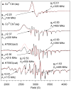
Experimental (solid line) and simulated (dotted line) X-Band (9.35 GHz) EPR spectra of: (a) aqueous Co2+Cbl, (b) aqueous Co2+Cbi+, (c) Co2+Cbl bound to H759G MetH(act), and (d) Co2+Cbl bound to H759G MetH(cat). Note that the EPR spectrum of aqueous Co2+Cbl was simulated with an isotropic A(N) coupling of 55 MHz to account for the additional fine structure. Complete parameter sets are available in Tables S1–S4.
Figure 8
Figure 8
Abs spectra of different forms of H759G MetH. When the as-isolated (i.e., Co2+Cbl-bound) H759G(act) form (solid trace) was reduced with titanium citrate, a Co1+Cbl-bound species (dotted trace) was obtained. Alternatively, when Co2+Cbl-bound H759G(act) was subjected to electrochemical methylation with AdoMet, a MeCbl-bound species was obtained (dashed trace). All spectra were obtained in 50 mM potassium phosphate buffer, pH 7.2, at 37° C.
Figure 9
Figure 9
Spectrophotometric determination of the Co2+Cbl/Co1+Cbl midpoint potential of H759G MetH. (A) H759G(act) was first photoreduced with 5-deazaflavin-3-sulfonate in the presence of the indicator dye methyl viologen to generate a Co1+Cbl-bound species (as evidenced by the decrease in absorbance at 468 nm), and spectra were then recorded to monitor the increase in absorbance at 468 nm associated with the spontaneous re-oxidation to the Co2+Cbl-bound H759G(act) form. (B) Nernst plot for the Co2+Cbl/Co1+Cbl reduction of H759G(act). From this plot, the midpoint potential was estimated to be −490 mV. (C) Attempts to photoreduce H759G(cat) with 5-deazaflavin-3-sulfonate in the presence of methyl viologen were unsuccessful, as evidenced by the lack of a change in absorbance at 468 nm right after photoreduction and over time as the indicator dye methyl viologen oxidized.
Figure 10
Figure 10
280 K Abs spectra of: (a) aqueous, base-on MeCbl, (b) aqueous, base-off MeCbl, and (c) H759G MetH(met). The vertical line serves to illustrate the successive blue-shift of the α-band from panels a through c.
Figure 11
Figure 11
7T, 280 K MCD spectra of: (a) aqueous, base-on MeCbl, (b) aqueous, base-off MeCbl, and (c) H759G MetH(met).
Figure 12
Figure 12
Space-filling representation of the X-ray crystal structure of the truncated H759G MetH variant (residues 649–1227) that only contains the Cbl- and AdoMet-binding modules and is thus forced to adopt the same conformation as H759G MetH(act) (14). Residues implicated in Fld docking are highlighted in white (3). Note that while the α-face of the Cbl cofactor is protected by the Cbl-binding module (red), the β-face is left partially solvent-accessible by the AdoMet-binding module (blue).
Scheme 1
Scheme 1

References

    1. Goulding CW, Postigo D, Matthews RG. Cobalamin-Dependent Methionine Syntahse Is a Modular Protein with Distinct Regions for Binding Homocysteine, Methyltetrahydrofolate, Cobalamin, and Adenosylmethionine. Biochemistry. 1997;36:8082–8091. - PubMed
    1. Drummond JT, Huang S, Blumenthal RM, Matthews RG. Assignment of Enzymatic Function to Specific Protein Regions of Cobalamin-Dependent Methionine Synthase from Escherichia coli. Biochemistry. 1993;32:9290–9295. - PubMed
    1. Hall DA, Jordan-Starck TC, Loo RO, Ludwig ML, Matthews RG. Interaction of Flavodoxin with Cobalamin-Dependent Methionine Synthase. Biochemistry. 2000;39:10711–10719. - PubMed
    1. Hall DA, Vander Kooi CW, Stasik CN, Stevens SY, Zuiderweg ERP. Mapping the Interaction Between Flavodoxin and its Physiological Partners Flavodoxin Reductase and Cobalamin-Dependent Methionine Synthase. Proc Natl Acad Sci U S A. 2001;98:9521–9526. - PMC - PubMed
    1. Evans JC, Huddler DP, Hilgers MT, Romanchuk G, Matthews RG, Ludwig ML. Structures of the N-terminal Modules Imply Large Domain Motions During Catalysis by Methionine Synthase. Proc Natl Acad Sci U S A. 2004;101:3729–3736. - PMC - PubMed

Publication types

MeSH terms

LinkOut - more resources