. 2007 Aug;9(8):1115-24.
doi: 10.1089/ars.2007.1674.

Aerobically derived lactate stimulates revascularization and tissue repair via redox mechanisms

Affiliations

Aerobically derived lactate stimulates revascularization and tissue repair via redox mechanisms

Thomas K Hunt et al. Antioxid Redox Signal. 2007 Aug.

Abstract

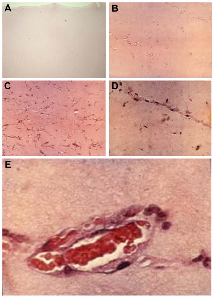
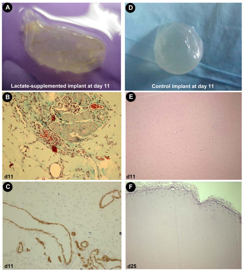
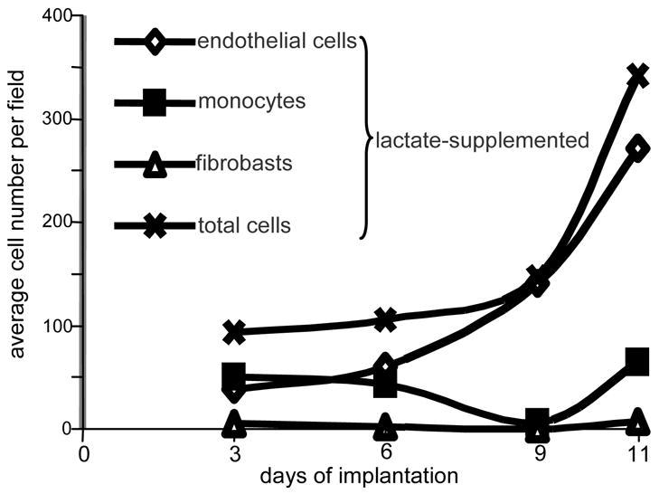
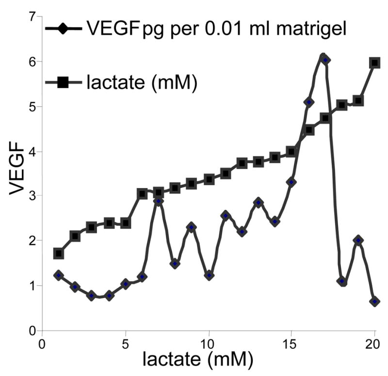
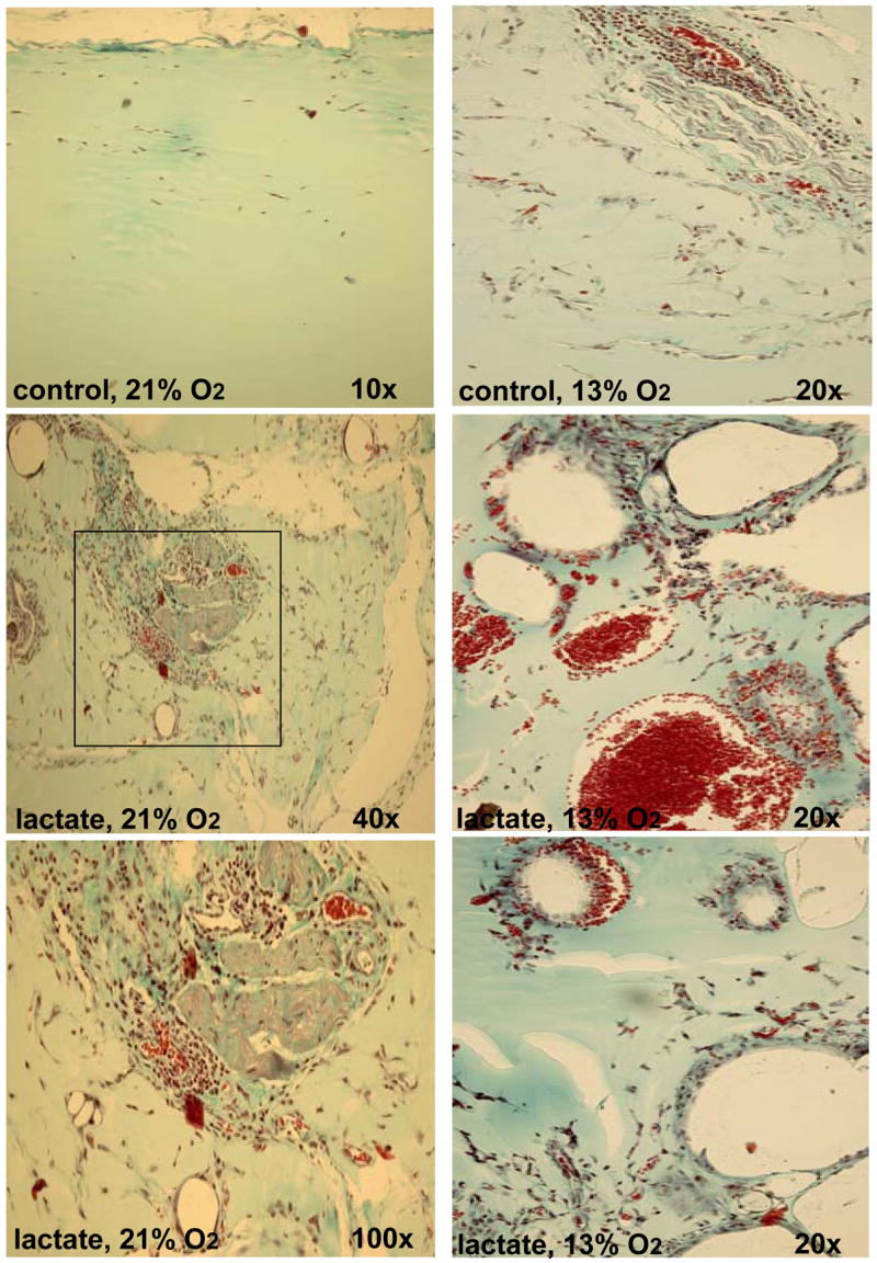
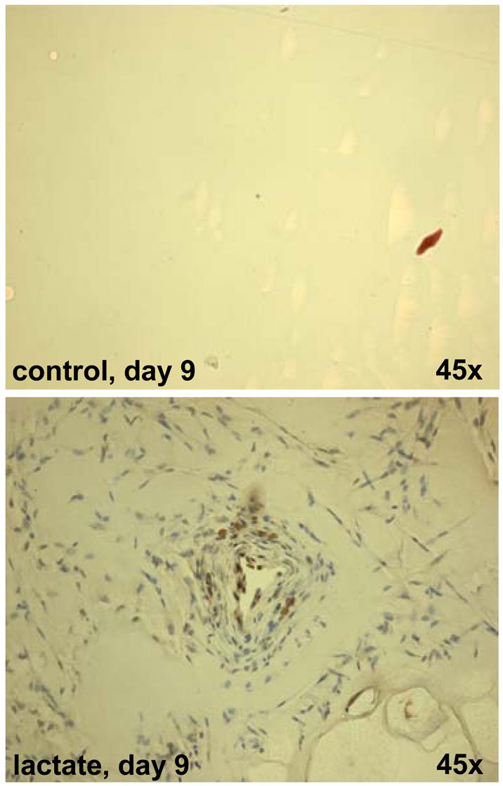
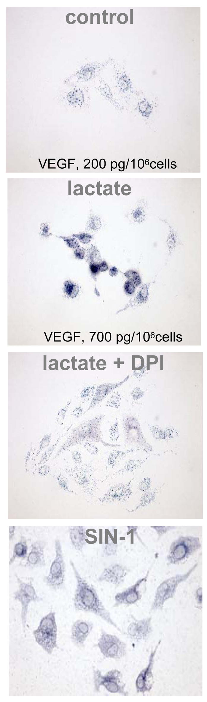
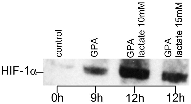
Hypoxia serves as a physiologic cue to drive an angiogenic response via HIF-dependent mechanisms. Interestingly, minor elevation of lactate levels in the tissue produces the same effect under aerobic conditions. Aerobic glycolysis contributes to lactate accumulation in the presence of oxygen, especially under inflammatory conditions. We previously postulated that aerobic lactate accumulation, already known to stimulate collagen deposition, will also stimulate angiogenesis. If substantiated, this concept would advance understanding of wound healing and aerobic angiogenesis because lactate accumulation has many aerobic sources. In this study, Matrigel plugs containing a powdered, hydrolyzable lactate polymer were implanted into the subcutaneous space of mice. Lactate monomer concentrations in the implant were consistent with wound levels for more than 11 days. They induced little inflammation but considerable VEGF production and were highly angiogenic, as opposed to controls. Arterial hypoxia abrogated angiogenesis. Furthermore, inhibition of lactate dehydrogenase by using oxamate also prevented the angiogenic effects of lactate. Lactate monomer, at concentrations found in cutaneous wounds, stabilized HIF-1alpha and increased VEGF levels in aerobically cultured human endothelial cells. Accumulated lactate, therefore, appears to convey the impression of "metabolic need" for vascularization, even in well-oxygenated and pH-neutral conditions. Lactate and oxygen together stimulate angiogenesis and matrix deposition.


Figures

Figure 1
Figure 1. Grading system used in assessing angiogenesis in Matrigel® implants
B , a score of 0 was given for acellular or rare cells; B, a score of 1 reflected scattered endothelial cells in small groups or linear arrangements but without lumens; C, a score of 2 represented endothelial cells in all quadrants of the section, prominent linear arrangements and some tube formation; D, a score of 3 was assigned for easily identified capillary tube formation, many containing red blood cells and small amounts of collagen; E, a score of 4 was reserved for larger vessels that accommodated more than 4 red cells abreast and multilayered vessels containing layers of collagen in vessel walls. Agreement between graders was >90%. In no case was the score-difference difference greater than 1 for the same observation by two different graders. The average of three repeated observations by the same graders was noted as the recordable score. Reproduced with permission from the publisher of Hopf et al. (17). For interpretation of the references to color in this figure legend, the reader is referred to the web version of this article at www.liebertonline.com/ars.
Figure 2
Figure 2. Blood vessel growth in lactate-supplemented Matrigel®
Matrigel® (n=80) containing hydrolysable poly-DL-lactide-co-glycolide (lactide:glycolide 50:50, mol wt 40,000–75,000) or no additive (control, n=100) were implanted subcutaneously, one injection in each flank just caudal to the ribcage. Four such lactate-supplemented implants were removed and examined microscopically at day 11 or 25 (control) as shown. A–C, lactate-supplemented (day 11); D–F, control (D&E, day 11; F, day 25). A&D, intact Matrigel® harvest; B&E, hematoxylin & eosin stain; C, CD31 immunostain. F, hematoxylin & eosin stain on day 25. For interpretation of the references to color in this figure legend, the reader is referred to the web version of this article at www.liebertonline.com/ars.
Figure 3
Figure 3. Kinetics of cellular infiltration in lactate-supplemented Matrigel®
Differential cell counts were done on the basis of cellular morphology as identified by hematoxylin & eosin stain. From each implant, two sections were examined. Four fields per slide were examined at 40x magnification avoiding fields in which the periphery of the implant could be seen. Control slides, from implants either non-treated or treated with high molecular weight lactide polymer, showed too few cells to enable differential scoring. Clearly, lactate attracted primarily monocytes and endothelial cells.
Figure 4
Figure 4. The relationship between lactate and VEGF levels in lactate-supplemented Matrigel®
Twenty implants, including both lactate-supplemented and controls were numbered in ascending order of magnitude of lactate as shown on the horizontal axis and the squared line. They were then matched with their corresponding VEGF level. When displayed in this manner, VEGF content with respect to lactate at 11 days appears to peak at about 6 mM lactate. However, the lactate concentrations are somewhat illusory in that the hydrolysis of the lactate powder is not linear with time. Thus they can give only an impression of what the concentrations were on days 1 to 11. Nevertheless, the data supports prior experience reported by Beckert (5) who found in cultured endothelial cells that VEGF concentration increased in response to lactate to a peak at about 15 mM and cells deteriorated above that level. This figure, therefore, indicates a significant relationship to increasing lactate in vivo that reaches a peak and then apparently deteriorates. Lactate levels in controls were in the range of 0.75 to 4 mM, the same range as found by others in blood and subcutaneous tissues.
Figure 5
Figure 5. Histological characterization of Matrigel® implants in normoxic and hypoxic mice
Thirty-six mice were each implanted with lactate-supplemented implant and one non-treated control. Twelve animals were held in each of three atmospheres: 13% (right column), 21% (left column), and 50% (not shown) oxygen for 11 days. A Pro-ox compact oxygen controller model 110 was used to maintain pO2 (Reming Bioinstruments, Redfield, NY). FiO2, humidity (60–80%), CO2 (<2 mm Hg) and temperature (23–25°C) were maintained within normal limits. Hypoxic (13%O2 ambience) were housed in a plexiglas chamber with a single gas inlet and outlet. Air was delivered at 1.0 L/min and nitrogen at 4.5 L/min from tanks connected to the chamber by thick-walled polyvinyl tubing. This reliably delivered 13.3 ± 1.3% oxygen to the chamber. Carbon dioxide levels were maintained at 0 mm Hg within the chamber with a CO2 absorber (Baralyme, Chemetron Medical Division, St. Louis, MO). A Datex Capnomac II [Datex Medical Instruments, Inc, Tewksbury, MA] was used to sample exhaust gas from the oxygen chambers to confirm the exposure pO2 and to ensure that CO2 did not accumulate. Compared to that in normoxic mice (left column), implants in hypoxic mice produced rare and large, thin-walled vessels. These photographs were selected to show as many vessels as possible and are not representative of their abundance. Compared to that in normoxic mice, small amounts of collagen were evident in implants in hypoxic mice. The sections were stained with Masson’s trichrome. For interpretation of the references to color in this figure legend, the reader is referred to the web version of this article at www.liebertonline.com/ars.
Figure 6
Figure 6. Recruitment of multipotent hematopoietic stem and progenitor cells in lactate-supplemented Matrigel®
CD117 staining is shown. CD117, also known as c-kit, steel factor receptor and stem cell factor receptor, encodes a 145 kD cell surface glycoprotein belonging to the class III receptor tyrosine kinase family. It is expressed on the majority of hematopoietic progenitor cells including multipotent hematopoietic stem cells as well as committed myeloid, erythroid, and lymphoid precursor cells. In addition to the potential for the differentiation of hematopoietic cells, CD117+ stem cells from murine bone marrow were reported to be capable of differentiation into smooth muscle cells, myocytes, and endothelial cells in vivo.1,2 CD117 is also expressed on few mature hematopoietic cells, e.g. mast cells. The sections were also richly populated with CD34+ cells (not shown). For interpretation of the references to color in this figure legend, the reader is referred to the web version of this article at www.liebertonline.com/ars.
Figure 7
Figure 7. Lactate-induced oxidant production in microvascular endothelial cells is likely to be NADPH oxidase dependent
. Human micro-vascular endothelial cells (HMVEC-d) purchased from Cambrex Bio Science (Walkersville, MD) and passages 5–9 were used for experiments. Cells were grown in endothelial basal medium 2 (CC-3156) containing EGM-2, MV (SingleQuots, Cambrex, Walkersville, MD, CC-4147), 100 units/ml penicillin, 100 units/ml streptomycin and 0,2 μg/ml amphotericin B. Cultures were maintained at 37° in humidified 95% air and 5% CO2 atmosphere. Cells were growth-arrested by incubating overnight in endothelial basal medium 2. Nitroblue tetrazolium (NBT) (1 mg/ml) was added followed by lactate (15 mM). pH was adjusted to 7.4. In similar experiments, effect of the NADPH oxidase inhibitor diphenylene iodonium (DPI, 10μM) and the oxidant donor SIN-1 (1 mM; 3-morpholino-syndnonimine) was tested. Note the differences in VEGF in response to lactate-supplementation.
Figure 8
Figure 8. Lactate-induced stabilization of HIF-1 α in oxygenated endothelial cells
. Endothelial cells were grown in 6 well plates to sub-confluent monolayers with a cell density of approximately 106 cells/well. After overnight serum starvation, cells were treated for nine hours with the prolyl-hydroxylase inhibitor GPA1734 (8,9-dihydroxy-7-methyl-benzo[b]quinolizinium bromide; kindly provided by Dr. M. Maragoudakis, Patras, Greece) and HIF-1α protein was detected by Western blotting. For immunoblotting, cells were washed with ice-cold PBS and collected by scraping into 1 ml PBS. After centrifugation at 1000 g for 5 min, pellets were homogenized with an equal volume of lysis buffer (20 mM Tris-HCl, pH 7.5 containing 150 mM NaCl) (Cell Signaling, Beverly, MA) containing protease inhibitors and incubated for 20 min on ice. After sonication for 15 seconds samples were subjected to centrifugation at 15,000 g for 20 min at 4 ° to separate membrane and cytosolic fractions, suspended in 2X SDS sample buffer (Cell Signaling, Beverly, MA) containing 1 mM DTT, and boiled for 5 minutes. Equal quantities of protein were separated by SDS-PAGE electrophoresis under reducing conditions using a 7.5% Tris-HCl gel (Bio-Rad, Hercules, CA). After transfer onto 0.45 μm nitrocellulose membranes (Millipore, Marlborough, MA), the membranes were blocked with 5% non-fat dry milk and probed with anti HIF-1α (1:1500; Novus Biologicals, Littleton, CO). GPA 1734 was used to block the proteosomal degradation pathway of HIF-1α.

References

    1. Albina JE, Mastrofrancesco B, Vessella JA, Louis CA, Henry WL, Jr, Reichner JS. HIF-1 expression in healing wounds: HIF-1alpha induction in primary inflammatory cells by TNF-alpha. Am J Physiol Cell Physiol. 2001;281:C1971–1977. - PubMed
    1. Ali MA, Yasui F, Matsugo S, Konishi T. The lactate-dependent enhancement of hydroxyl radical generation by the Fenton reaction. Free Radic Res. 2000;32:429–438. - PubMed
    1. Allen DB, Maguire JJ, Mahdavian M, Wicke C, Marcocci L, Scheuenstuhl H, Chang M, Le AX, Hopf HW, Hunt TK. Wound hypoxia and acidosis limit neutrophil bacterial killing mechanisms. Arch Surg. 1997;132:991–996. - PubMed
    1. Attard JA, Raval MJ, Martin GR, Kolb J, Afrouzian M, Buie WD, Sigalet DL. The effects of systemic hypoxia on colon anastomotic healing: an animal model. Dis Colon Rectum. 2005;48:1460–1470. - PubMed
    1. Beckert S, Farrahi F, Aslam RS, Scheuenstuhl H, Konigsrainer A, Hussain MZ, Hunt TK. Lactate stimulates endothelial cell migration. Wound Repair Regen. 2006;14:321–324. - PubMed

Publication types

LinkOut - more resources