. 2007 Aug 31;282(35):25668-76.
doi: 10.1074/jbc.M702076200. Epub 2007 Jun 13.

Multiple and additive functions of ALDH3A1 and ALDH1A1: cataract phenotype and ocular oxidative damage in Aldh3a1(-/-)/Aldh1a1(-/-) knock-out mice

Affiliations

Multiple and additive functions of ALDH3A1 and ALDH1A1: cataract phenotype and ocular oxidative damage in Aldh3a1(-/-)/Aldh1a1(-/-) knock-out mice

Natalie Lassen et al. J Biol Chem. .

Abstract

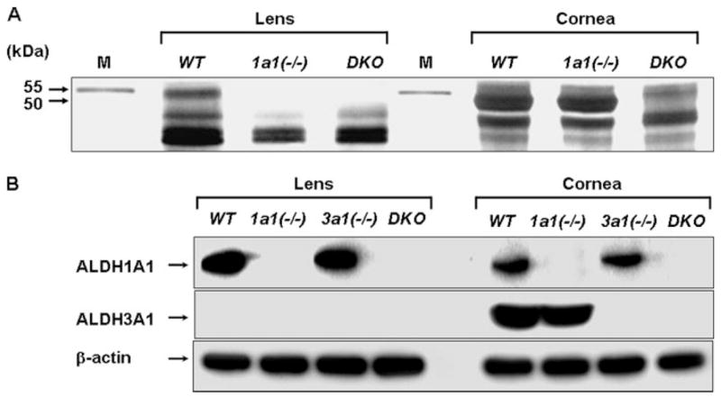
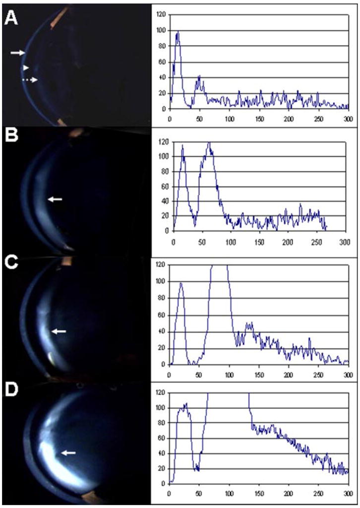
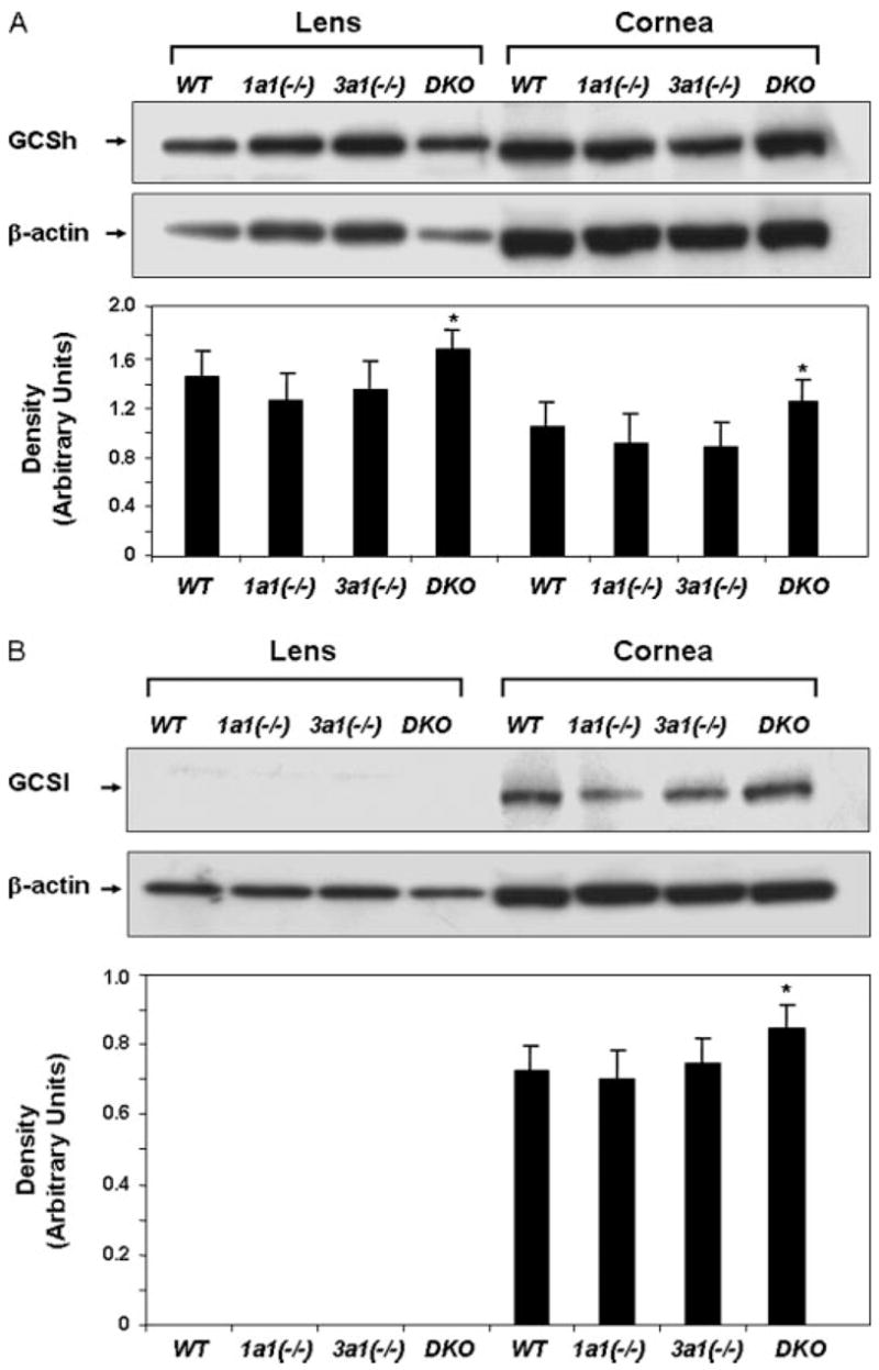
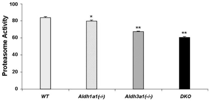
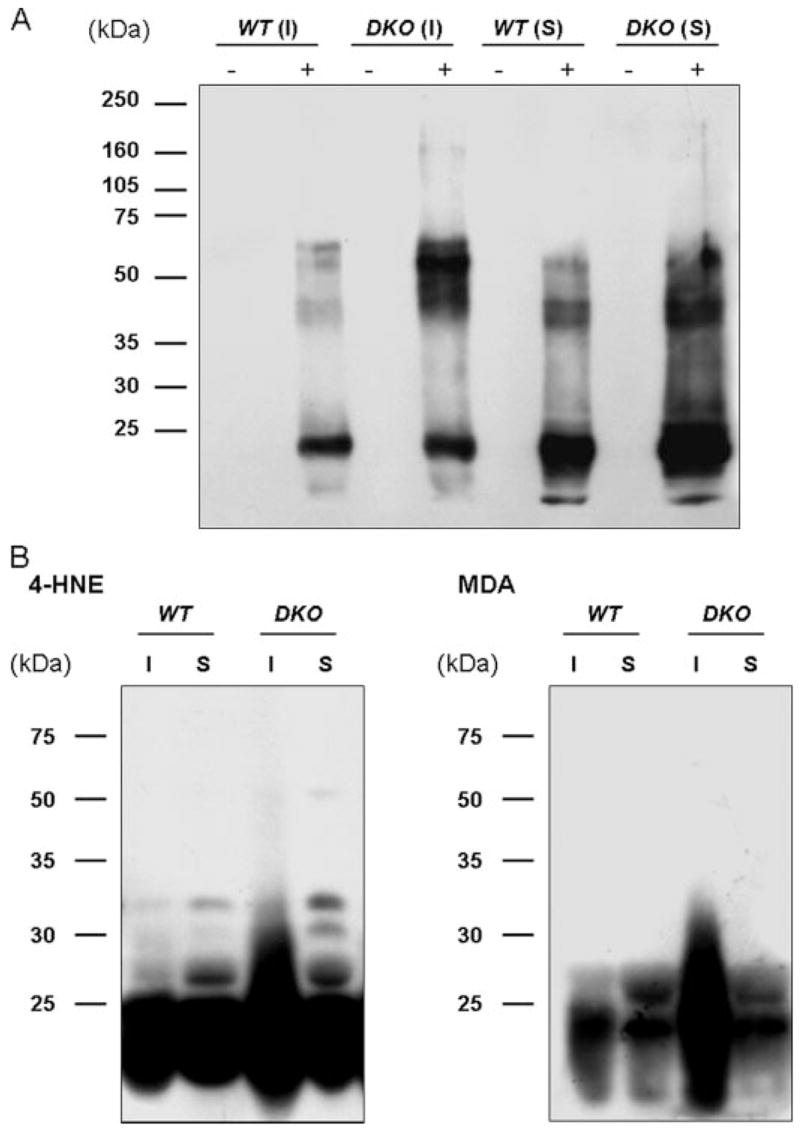
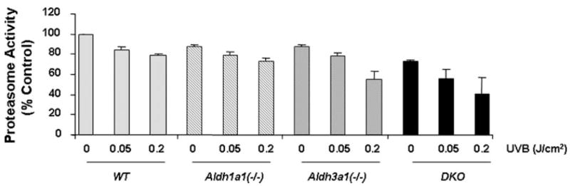
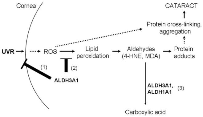
ALDH3A1 (aldehyde dehydrogenase 3A1) is abundant in the mouse cornea but undetectable in the lens, and ALDH1A1 is present at lower (catalytic) levels in the cornea and lens. To test the hypothesis that ALDH3A1 and ALDH1A1 protect the anterior segment of the eye against environmentally induced oxidative damage, Aldh1a1(-/-)/Aldh3a1(-/-) double knock-out and Aldh1a1(-/-) and Aldh3a1(-/-) single knock-out mice were evaluated for biochemical changes and cataract formation (lens opacification). The Aldh1a1/Aldh3a1- and Aldh3a1-null mice develop cataracts in the anterior and posterior subcapsular regions as well as punctate opacities in the cortex by 1 month of age. The Aldh1a1-null mice also develop cataracts later in life (6-9 months of age). One- to three-month-old Aldh-null mice exposed to UVB exhibited accelerated anterior lens subcapsular opacification, which was more pronounced in Aldh3a1(-/-) and Aldh3a1(-/-)/Aldh1a1(-/-) mice compared with Aldh1a1(-/-) and wild type animals. Cataract formation was associated with decreased proteasomal activity, increased protein oxidation, increased GSH levels, and increased levels of 4-hydroxy-2-nonenal- and malondialdehyde-protein adducts. In conclusion, these findings support the hypothesis that corneal ALDH3A1 and lens ALDH1A1 protect the eye against cataract formation via nonenzymatic (light filtering) and enzymatic (detoxification) functions.


Figures

FIGURE 1
FIGURE 1. Detection of ALDH1A1 and ALDH3A1 proteins in lens and cornea
A, SDS-polyacrylamide gel with silver staining of proteins (10 μg) extracted from lens and cornea from wild type (WT), Aldh1a1(−/−) (1a1(−/−)), and double knock-out mice (DKO). The positions of the protein bands corresponding to ALDH1A1 and ALDH3A1 are indicated by the arrows (55 and 50 kDa, respectively). M, molecular weight markers. B, Western blot analyses of ALDH1A1 and ALDH3A1 proteins (20 μg) in wild type, Aldh1a1(−/−), and Aldh3a1(−/−) (3a1(−/−)) single and double knock-out mice. β-Actin expression was used to show equal loading.
FIGURE 2
FIGURE 2. Slit lamp biomicroscopy and corresponding densitometry tracings of the anterior capsule and cortex of wild type, Aldh1a1(−/−), and Aldh3a1(−/−) single and double knock-out mice, 6 months of age
The left light band is the cornea (arrow). The adjacent dark region is the water-filled anterior chamber (arrowhead). The next light band is the anterior capsule and lens epithelium (dashed arrow), and the remainder of the image is the central lens. Densitometry scans of pixel intensity through the center of each image are shown on the right side of the slit lamp photographs with cornea peaks normalized to an arbitrary value of 100, assuming that all images had the same corneal clarity. A, wild type. The lens is clear with a relatively flat graph after the peak due to the capsule and epithelium. B, Aldh1a1(−/−). There is mild scattering from the anterior subcapsular cataract (arrow). C, Aldh3a1(−/−). There is denser anterior subcapsular cataract (arrow). D, Aldh1a1(−/−)/Aldh3a1(−/−). There is definite and denser anterior subcapsular cataract (arrow).
FIGURE 3
FIGURE 3. Increased GCS expression in the lens and cornea of the double knock-out mice (6 months of age)
Shown is Western blot analysis of GCSh (73 kDa) (A) and GCSl (30 kDa) (B) subunits in lens and cornea lysates. Thirty μg of cellular protein was loaded per lane, and equal loading was confirmed by probing the membranes with β-actin antibody. S.E. was less than 10% in all cases. *, p < 0.05, Student’s unpaired t test, compared with respective wild type control mice.
FIGURE 4
FIGURE 4. Decrease of chymotrypsin-like proteasome activity in Aldh1a1(−/−)/Aldh3a1(−/−) double knock-out mice
Lenses from wild type, Aldh1a1(−/−), and Aldh3a1(−/−) single and double knock-out mice, 6 months of age, were extracted, and fresh lysates were processed for chymotrypsin-like proteasome activity. The activities are expressed as the rate of substrate conversion/min × mg of protein. Values represent mean ± S.E. (n = 3/group). An overall significant difference in proteasome activity was observed among the different genotypes (F3,8 = 264.2, p < 0.001). Post hoc Bonferroni’s t test indicated that all groups were significantly different from each other. *, p < 0.05 comparing Aldh1a1(−/−) to wild type and p < 0.001 comparing Aldh1a1(−/−) to either Aldh3a1(−/−) or double knock-out mice. **, p < 0.001 comparing the designated group to any other group.
FIGURE 5
FIGURE 5. Detection of oxidized proteins (A) and 4-HNE- and MDA-adducted proteins (B) in lens extracts from wild type and Aldh1a1(−/−)/Aldh3a1(−/−) double knock-out mice
Lenses from wild type (WT) and double knock-out (DKO) mice, 6 months of age, were separated into soluble (S) and insoluble (I) fractions. A, 2,4-dinitrophenylhydrazine-derivatized (+) and nonderivatized (−) proteins were separated by SDS-PAGE (10%) and transferred to polyvinylidene difluoride membranes. Oxidized proteins were recognized by an antibody specific to the 2,4-dinitrophenylhydrazone-derivatized residues and detected by enhanced chemiluminescent reagents. B, equal amounts of lens homogenates were subjected to SDS-PAGE (10%) and transferred to polyvinylidene difluoride membranes. 4-HNE- and MDA-adducted proteins were detected by Western blotting. Protein molecular mass (kDa) markers are given on the left.
FIGURE 6
FIGURE 6. Decrease of chymotrypsin-like proteasome activity in Aldh genetic stocks after UVB exposure
Wild type, Aldh1a1(−/−), and Aldh3a1(−/−) single and Aldh1a1(−/−)/Aldh3a1(−/−) double knock-out mice, 1–3 months of age, were exposed to two different doses of UVB light (0.05 and 0.2 J/cm2). The lenses were subsequently extracted and processed for chymotrypsin-like proteasome activity. The activities were normalized to percentage of control. Values represent mean ± S.E. (n = 3/group).
SCHEME 1
SCHEME 1. Possible mechanisms by which ALDH3A1 and ALDH1A1 protect the eye from UV-induced cataract formation
1, ALDH3A1 in the cornea directly absorbs UV light, acting as a UV filter. 2, ALDH3A1 potentially acts as a ROS scavenger, thereby preventing protein cross-linking and aggregation. 3, combined role of ALDH1A1 and ALDH3A1 in detoxification of 4-HNE and MDA derived from lipid peroxidation of cellular membranes, protecting ocular tissues from protein cross-linking and aggregation, which would lead to proteasome inhibition and eventually cataract formation.

References

    1. Ringvold A. Acta Ophthalmol Scand. 1998;76:149–153. - PubMed
    1. Spector A. FASEB J. 1995;9:1173–1182. - PubMed
    1. Spector A. J Ocul Pharmacol Ther. 2000;16:193–201. - PubMed
    1. Shinohara T, Singh DP, Chylack LT., Jr J Ocul Pharmacol Ther. 2000;16:181–191. - PubMed
    1. Cruickshanks KJ, Klein BE, Klein R. Am J Public Health. 1992;82:1658–1662. - PMC - PubMed

Publication types

MeSH terms