. 2007;35(12):4076-85.
doi: 10.1093/nar/gkm434. Epub 2007 Jun 12.

Nuclear interactions of topoisomerase II alpha and beta with phospholipid scramblase 1

Affiliations

Nuclear interactions of topoisomerase II alpha and beta with phospholipid scramblase 1

Jessica P Wyles et al. Nucleic Acids Res. 2007.

Abstract

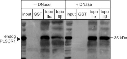
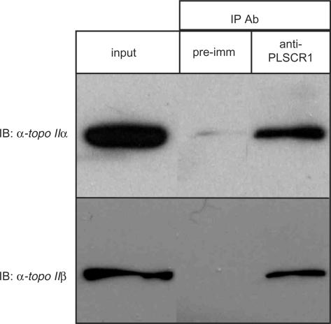
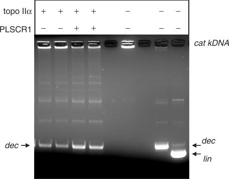
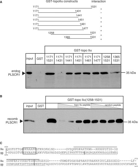
DNA topoisomerase (topo) II modulates DNA topology and is essential for cell division. There are two isoforms of topo II (alpha and beta) that have limited functional redundancy, although their catalytic mechanisms appear the same. Using their COOH-terminal domains (CTDs) in yeast two-hybrid analysis, we have identified phospholipid scramblase 1 (PLSCR1) as a binding partner of both topo II alpha and beta. Although predominantly a plasma membrane protein involved in phosphatidylserine externalization, PLSCR1 can also be imported into the nucleus where it may have a tumour suppressor function. The interactions of PLSCR1 and topo II were confirmed by pull-down assays with topo II alpha and beta CTD fusion proteins and endogenous PLSCR1, and by co-immunoprecipitation of endogenous PLSCR1 and topo II alpha and beta from HeLa cell nuclear extracts. PLSCR1 also increased the decatenation activity of human topo IIalpha. A conserved basic sequence in the CTD of topo IIalpha was identified as being essential for binding to PLSCR1 and binding of the two proteins could be inhibited by a synthetic peptide corresponding to topo IIalpha amino acids 1430-1441. These studies reveal for the first time a physical and functional interaction between topo II and PLSCR1.


Figures

Figure 1.
Figure 1.
GST-tagged topo IIα CTD and topo IIβ CTD interact with PLSCR1. An NP-40 soluble HeLa cell lysate (150 μg protein) was incubated sequentially with DNase1 (20 U), GST-tagged topo IIα or topo IIβ CTD fusion proteins (5 μg) and GSH-Sepharose beads (25 μl of 50% slurry) as described in Materials and Methods section. The beads were collected by centrifugation and bound proteins analysed by immunoblotting with rabbit PLSCR1 antiserum. Input lane represents 5% of lysate used in binding assay.
Figure 2.
Figure 2.
PLSCR1 co-fractionates with topo II α and β. (A) Nuclear and NP-40 soluble lysates were prepared from HeLa cells as described in Materials and Methods section for co-immunoprecipitation and GST-pull-down assays, respectively. Samples representing equivalent numbers of cells were analysed by immunoblotting with antibodies against PLSCR1, topo IIα (mAb 8D2) and topo IIβ (mAb 3H10). (B) The same samples described in Panel A were analysed for calnexin, LAMP2 and tubulin content by immunoblotting to confirm the absence of contaminating ER, lysosomal and cytoskeletal proteins, respectively. To prevent overexposure of the film due to high intensity signals, the analyses for tubulin and calnexin content used 10% of the number of cells used for other immunoblots. Nuc, nuclear lysate; NP-40, NP-40 soluble lysate; TX-100, Triton X-100 soluble lysate.
Figure 3.
Figure 3.
Endogenous topo II α and β co-immunoprecipitate with endogenous PLSCR1. Nuclear extracts prepared from HeLa cells were subjected to co-immunoprecipitation with an anti-PLSCR1 antiserum or pre-immune serum as described in Materials and Methods section. Immunoprecipitated samples were then analysed by immunoblotting with topo II α and β specific mAbs 8D2 and 3H10, respectively. Input lanes represent 5% of sample used in the assay. Shown are results from a representative experiment and similar results were obtained in two additional experiments.
Figure 4.
Figure 4.
Colocalization of endogenous topo IIα and PLSCR1. HeLa cells were cultured on gelatinized glass coverslips, fixed in formaldehyde and permeabilized with Triton X-100. Topo IIα was detected with mAb 8D2 in conjunction with Alexa546 ™ -conjugated goat anti-mouse secondary antibody (Panels A and D, red signal) and PLSCR1 was detected with rabbit PLSCR1 antiserum or the pre-immune serum (Panels B and E, respectively, green signal) in conjunction with Alexa488 ™ -conjugated goat anti-rabbit secondary antibody. Merged images are shown in Panels C (A + B) and F (D + E).
Figure 5.
Figure 5.
PLSCR1 stimulates the decatenation activity of topo IIα. Topo II decatenating activity was assayed using purified topo IIα enzyme in the presence and absence of untagged recombinant PLSCR1 as described in Materials and Methods section. Similar results to those shown were obtained in two additional experiments. The positions of the catenated kDNA (cat kDNA), decatenated kDNA minicircles (dec) and linear (lin) kDNA are indicated.
Figure 6.
Figure 6.
Localization of PLSCR1 binding to amino acids 1432-1441 of topo IIα. (A) Upper panel, Schematic diagram showing the eight GST-tagged topo IIαCTD fusion proteins used to define the region of topo IIα involved in binding to PLSCR1. Lower panel, NP-40 solubilized HeLa cell lysates were incubated with each of the eight GST-topo IIαCTD fusion proteins and complexes pulled down with GSH-Sepharose beads as described in Materials and Methods section. The proteins were eluted and immunoblotted with PLSCR1 antiserum. (B) Untagged recombinant PLSCR1 was incubated with increasing concentrations of synthetic dodecapeptides corresponding to topo IIα residues 1430-1441 or as a control, randomly scrambled topo IIα residues 1430-1441, and GST-topo IIα1258-1531 or GST alone (control). Complexes were pulled down with GSH-Sepharose beads and bound proteins analysed by immunoblotting with PLSCR1 antiserum. The experiment shown was repeated three or more times with comparable results. (C) The sequences of the COOH-termini of topo IIα and topo IIβ were aligned using ClustalW. The basic conserved putative PLSCR1 binding motifs in topo IIα and topo IIβ are boxed; the vertical bars in the topo II sequences represent intron/exon boundaries (43), and the functional NLS sequences in this region are underlined (topo IIα, solid line; topo IIβ, broken line) (24).

References

    1. Wang JC. Cellular roles of DNA topoisomerases: a molecular perspective. Nat. Rev. Mol. Cell Biol. 2002;3:430–440. - PubMed
    1. Woessner RD, Mattern MR, Mirabelli CK, Johnson RK, Drake FH. Proliferation- and cell cycle-dependent differences in expression of the 170 kilodalton and 180 kilodalton forms of topoisomerase II in NIH-3T3 cells. Cell Growth Differ. 1991;2:209–214. - PubMed
    1. Bauman ME, Holden JA, Brown KA, Harker WG, Perkins SL. Differential immunohistochemical staining for DNA topoisomerase II α and β in human tissues and for DNA topoisomerase IIβ in non-Hodgkin's lymphomas. Mod. Pathol. 1997;10:168–175. - PubMed
    1. Watanabe M, Tsutsui K, Inoue Y. Differential expressions of the topoisomerase IIα and IIβ mRNAs in developing rat brain. Neurosci. Res. 1994;19:51–57. - PubMed
    1. Tsutsui K, Sano K, Kikuchi A, Tokunaga A. Involvement of DNA topoisomerase IIβ in neuronal differentiation. J. Biol. Chem. 2001;276:5769–5778. - PubMed

Publication types

MeSH terms