

eF-seek: prediction of the functional sites of proteins by searching for similar electrostatic potential and molecular surface shape
- PMID: 17567616
- PMCID: PMC1933152
- DOI: 10.1093/nar/gkm351
eF-seek: prediction of the functional sites of proteins by searching for similar electrostatic potential and molecular surface shape
Abstract
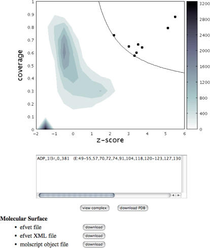
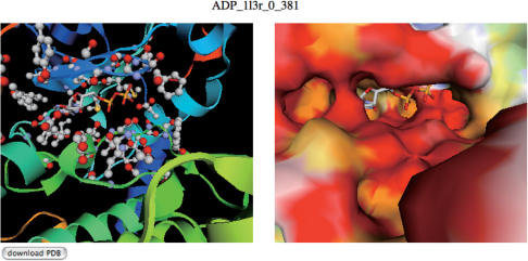
We have developed a method to predict ligand-binding sites in a new protein structure by searching for similar binding sites in the Protein Data Bank (PDB). The similarities are measured according to the shapes of the molecular surfaces and their electrostatic potentials. A new web server, eF-seek, provides an interface to our search method. It simply requires a coordinate file in the PDB format, and generates a prediction result as a virtual complex structure, with the putative ligands in a PDB format file as the output. In addition, the predicted interacting interface is displayed to facilitate the examination of the virtual complex structure on our own applet viewer with the web browser (URL: http://eF-site.hgc.jp/eF-seek).
Figures
References
-
- Bateman A, Valencia A. Structural genomics meets computational biology. Bioinformatics. 2006;22:2319. - PubMed
-
- Lundstrom K. Structural genomics: the ultimate approach for rational drug design. Mol. Biotechnol. 2006;34:205–212. - PubMed
-
- Rigden DJ. Understanding the cell in terms of structure and function: insights from structural genomics. Curr. Opin. Biotechnol. 2006;17:457–464. - PubMed
-
- Chandonia JM, Brenner SE. The impact of structural genomics: expectations and outcomes. Science. 2006;311:347–351. - PubMed
-
- Watson JD, Laskowski RA, Thornton JM. Predicting protein function from sequence and structural data. Curr. Opin. Struct. Biol. 2005;15:275–284. - PubMed
