. 2007 Sep;81(17):9152-61.
doi: 10.1128/JVI.00888-07. Epub 2007 Jun 13.

Fusion promotion by a paramyxovirus hemagglutinin-neuraminidase protein: pH modulation of receptor avidity of binding sites I and II

Affiliations

Fusion promotion by a paramyxovirus hemagglutinin-neuraminidase protein: pH modulation of receptor avidity of binding sites I and II

Laura M Palermo et al. J Virol. 2007 Sep.

Abstract

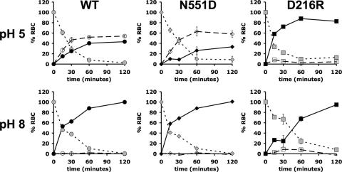
Paramyxoviruses, including the childhood respiratory pathogen human parainfluenza virus type 3 (HPIV3), possess an envelope protein hemagglutinin-neuraminidase (HN) that has receptor-cleaving (neuraminidase), as well as receptor-binding, activity. HN is a type II transmembrane glycoprotein, present on the surface of the virus as a tetramer composed of two dimers. HN is also essential for activating the fusion protein (F) to mediate merger of the viral envelope with the host cell membrane. This initial step of viral entry occurs at the host cell surface at neutral pH. The HN molecule carries out these three different critical activities at specific points in the process of viral entry, and understanding the regulation of these activities is key for the design of strategies that block infection. One bifunctional site (site I) on the HN of HPIV3 possesses both receptor binding and neuraminidase activities, and we recently obtained experimental evidence for a second receptor binding site (site II) on HPIV3 HN. Mutation of HN at specific residues at this site, which is next to the HN dimer interface, confers enhanced fusion properties, without affecting neuraminidase activity or receptor binding at neutral pH. We now demonstrate that mutations at this site II, as well as at site I, confer pH dependence on HN's receptor avidity. These mutations permit pH to modulate the binding and fusion processes of the virus, potentially providing regulation at specific stages of the viral life cycle.


Figures

FIG. 1.
FIG. 1.
Quantitation of the effect of temperature and neuraminidase pretreatment on HAD by cells expressing D216R or WT HN. HAD assays at 4°C (A) and 37°C (B), without (▪) and after (□) neuraminidase pretreatment, were carried out as described in Materials and Methods. The bars (absorbance at 405 nm) represent means of results ± the standard deviation (SD) on triplicate cell monolayers from a representative experiment.
FIG. 2.
FIG. 2.
Sensitivity of receptor-binding activity of WT HN, N551D HN, and D216R HN to pH. Cell monolayers transiently expressing the indicated HNs were assayed by HAD (at 4°C) using a series of receptor-depleted RBCs as described in Materials and Methods. The binding (y axis) at the indicated degree of receptor depletion, expressed as milliunits of neuraminidase treatment (x axis) is expressed as the percentage of binding obtained with undepleted RBCs at pH 5 (open symbols, solid lines) and pH 8 (black symbols, dashed lines). The results are means ± the SD of results on triplicate wells from a representative experiment.
FIG. 3.
FIG. 3.
Dependence of F activation by expressed WT and variant HNs on pH: quantitation of the amount of RBCs that are released, bound, or fused via F triggering at pH 5.0 or 8.0. For the F-activation assay, monolayers of cells coexpressing WT F and WT HN, D216R HN, or N551D HN with RBCs bound to them at 4°C were transferred to 37°C. At the indicated time points of incubation, RBCs that were (i) released by the HNs’ neuraminidase (open symbols, dashed lines), (ii) bound (gray symbols, dotted line), and (iii) fused (black symbols, solid black line) were quantified. The results are means ± the SD of results on triplicate wells from a representative experiment.
FIG. 4.
FIG. 4.
Dependence of receptor release by expressed WT and variant HNs on pH: release of target RBCs that were prebound to WT HN-, N551D HN-, and D216R HN-expressing cells at pH 5.0 or pH 8.0. Monolayers of cells coexpressing a cleavage site mutant F (K108G) and WT HN, D216R HN, or N551D HN with RBCs bound to them at 4°C were transferred to 37°C. After various times of incubation, RBCs that were released by the HNs’ neuraminidase at pH 5.0 (open symbols, solid lines) and pH 8.0 (black symbols, dashed lines) were quantified. The results are means ± the SD of results on triplicate wells from a representative experiment.
FIG. 5.
FIG. 5.
Modulation by pH of F activation by WT and variant HNs. Monolayers of cells coexpressing WT F with WT HN, D216R HN, or N551D HN with receptor-depleted RBCs bound to them at 4°C were transferred to 37°C. RBC fusion at pH 5.0 and 8.0 was quantitated as in Fig. 2. The extent of RBC fusion is expressed (y axis) as a percentage of total RBCs bound at each RBC depletion level. Bars: ▪, WT HN; □, N551D HN; □, D216R HN. The results are means ± the SD of results on triplicate wells from a representative experiment.
FIG. 6.
FIG. 6.
Cell-cell fusion mediated by WT F coexpressed with WT HN, D216R HN, or N551D HN. HPIV3 HN/F-coexpressing cells were allowed to fuse with receptor bearing cells at 37°C at pH 5.0, 7.3, or 8.0. Fusion was quantitated by using a β-Gal complementation assay as described in Materials and Methods. Cell fusion (y axis) in the presence of each coexpressed variant HN (indicated on the x axis) is expressed as the percentage of the fusion in the presence of WT HN at each pH. The bars represent means ± the SD of results from three different experiments.
FIG. 7.
FIG. 7.
Modulation of viral entry by pH. Cell monolayers treated with increasing amounts of neuraminidase were infected with viruses bearing WT or N551D HN at pH 5.0 or 8.0, and viral entry was quantitated by plaque assay. Infection (y axis) is expressed as the percentage of infection obtained on untreated cells at each pH. The bars represent means ± the SD of results from three different experiments.
FIG. 8.
FIG. 8.
Schematic structure of the HPIV3 HN dimer. Ribbon diagram showing the location of residue D216 (red) and N551 (green) in each monomer. Site I, neuraminidase activity and 1° receptor binding; site II, 2° receptor binding and F activation.

Similar articles

Cited by

References

    1. Bissonnette, M. L., S. A. Connolly, D. F. Young, R. E. Randall, R. G. Paterson, and R. A. Lamb. 2006. Analysis of the pH requirement for membrane fusion of different isolates of the paramyxovirus parainfluenza virus 5. J. Virol. 80:3071-3077. - PMC - PubMed
    1. Bousse, T., and T. Takimoto. 2006. Mutation at residue 523 creates a second receptor binding site on human parainfluenza virus type 1 hemagglutinin-neuraminidase protein. J. Virol. 80:9009-9016. - PMC - PubMed
    1. Crennell, S., T. Takimoto, A. Portner, and G. Taylor. 2000. Crystal structure of the multifunctional paramyxovirus hemagglutinin-neuraminidase. Nat. Struct. Biol. 7:1068-1074. - PubMed
    1. Deng, R., Z. Wang, P. J. Mahon, M. Marinello, A. Mirza, and R. M. Iorio. 1999. Mutations in the Newcastle disease virus hemagglutinin-neuraminidase protein that interfere with its ability to interact with the homologous F protein in the promotion of fusion. Virology 253:43-54. - PubMed
    1. Deng, R., Z. Wang, A. Mirza, and R. Iorio. 1995. Localization of a domain on the paramyxovirus attachment protein required for the promotion of cellular fusion by its homologous fusion protein spike. Virology 209:457-469. - PubMed

Publication types

LinkOut - more resources