. 2007 Sep;81(17):8933-43.
doi: 10.1128/JVI.00878-07. Epub 2007 Jun 13.

Functionally distinct transmission of human immunodeficiency virus type 1 mediated by immature and mature dendritic cells

Affiliations

Functionally distinct transmission of human immunodeficiency virus type 1 mediated by immature and mature dendritic cells

Jian-Hua Wang et al. J Virol. 2007 Sep.

Abstract

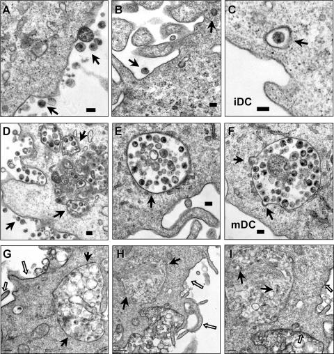
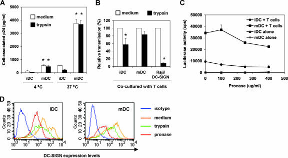
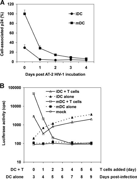
Dendritic cells (DCs) potently stimulate the transmission of human immunodeficiency virus type 1 (HIV-1) to CD4(+) T cells. Immature DCs (iDCs) located in submucosal tissues can capture HIV-1 and migrate to lymphoid tissues, where they become mature DCs (mDCs) for effective antigen presentation. DC maturation promotes HIV-1 transmission; however, the underlying mechanisms remain unclear. Here we have compared monocyte-derived iDCs and mDCs for their efficiencies and mechanisms of HIV-1 transmission. We have found that mDCs significantly facilitate HIV-1 endocytosis and efficiently concentrate HIV-1 at virological synapses, which contributes to mDC-enhanced viral transmission, at least in part. mDCs were more efficient than iDCs in transferring HIV-1 to various types of target cells independently of C-type lectins, which partially accounted for iDC-mediated HIV-1 transmission. Efficient HIV-1 trans-infection mediated by iDCs and mDCs required contact between DCs and target cells. Moreover, rapid HIV-1 degradation occurred in both iDCs and mDCs, which correlated with the lack of HIV-1 retention-mediated long-term viral transmission. Our results provide new insights into the mechanisms underlying DC-mediated HIV-1 transmission, suggesting that HIV-1 exploits mDCs to facilitate its dissemination within lymphoid tissues.


Figures

FIG. 1.
FIG. 1.
Mature DCs enhance HIV transmission to different types of target cells independently of C-type lectins. (A) Surface markers of iDCs and mDCs. Monocyte-derived iDCs were cultured with LPS to generate mDCs. Cell surface markers were stained with specific MAbs or isotype-matched IgG controls and analyzed by flow cytometry. The histogram peaks of CD11c staining on iDCs and mDCs were overlapped. (B) Enhanced HIV transmission by mDCs is independent of C-type lectins. DCs and Raji/DC-SIGN cells were preincubated separately with medium, anti-DC-SIGN cocktails, and mannan prior to HIV incubation, as described previously (57). Raji/DC-SIGN cells, iDCs, and mDCs were pulsed separately with single-cycle, luciferase reporter R5-tropic HIV-Luc/JRFL (multiplicity of infection [MOI], 0.2), washed, and cocultured separately with autologous PBLs, the CD4+ T-cell line Hut/CCR5, and the HIV indicator cells GHOST/R5. HIV infection was determined after 2 days by measuring the luciferase activity. (C) No detectable HIV cis-infection in DCs and Raji/DC-SIGN cells. Cells were infected with HIV-Luc/JRFL (MOI, 0.2), and viral infection was determined at 2 dpi. (D) mDCs enhance transmission of HIV pseudotyped with X4-tropic Env (HXB2). Transmission of HIV-Luc/HXB2 with DCs as donors and Hut/CCR5 cells as targets was performed as described for panel B. The asterisk indicates a significant difference (P < 0.05) compared with iDCs. The data show the means ± standard deviations of triplicate samples. One representative experiment out of four is shown. cps, counts per second.
FIG. 2.
FIG. 2.
DC-target cell contact is required for efficient HIV transmission mediated by iDCs and mDCs. Transmission of HIV-Luc/JRFL with iDCs and mDCs as donors and (A) Hut/CCR5 cells or (B) GHOST/R5 cells as targets was performed as described in the legend to Fig. 1B. Transwell culture plates with membrane pore sizes of 3 μm were used to separate DCs and target cells (Transwell). HIV infection was determined after 2 dpi by measuring the luciferase activity. The data show the means ± standard deviations of triplicate samples. One representative experiment out of three is shown. cps, counts per second.
FIG. 3.
FIG. 3.
Enhanced HIV endocytosis and distinct viral trafficking in mDCs relative to iDCs. DCs were exposed to AT-2-inactivated R5 HIV for 1.5 h, washed thoroughly, fixed, and prepared for electron microscopy. (A, B, and C) Cell surface-bound HIV and internalized viral particles in iDCs. (D, E, and F) HIV internalization is significantly enhanced in mDCs. Arrows indicate DC surface-associated HIV particles or intracellular compartments that trapped intact HIV particles. (G, H, and I) RR labeling of mDC plasma membranes. (I) Higher-magnification image of panel H (a partial area). The open arrows indicate RR-labeled mDC plasma membranes, and the black arrows point to HIV-containing compartments and HIV particles that were not labeled with RR. Scale bars, 0.1 μm (A to F), 0.2 μm (G, I), and 0.5 μm (H).
FIG. 4.
FIG. 4.
mDCs are more potent than iDCs in protecting HIV from proteolysis. (A) mDCs enhance HIV binding and internalization. DCs were incubated with HIV-Luc/JRFL (30 ng of p24) at 4°C or 37°C for 2 h, washed and treated with trypsin or medium, and then lysed for HIV p24 quantification. Asterisks indicate significant differences (P < 0.01) compared with iDCs at the same temperature. (B) DCs protect captured HIV from trypsin cleavage. HIV-pulsed iDCs, mDCs, and Raji/DC-SIGN cells were separately treated with trypsin before coculture with Hut/CCR5 target cells. Transmission of HIV-Luc/JRFL to Hut/CCR5 target cells was performed as described in the legend to Fig. 1B. The average results of four independent experiments are shown. Values for medium controls were set at 100%. Asterisks indicate significant differences (P < 0.05) between trypsin-treated samples and medium controls. (C) DCs protect captured HIV from pronase cleavage. HIV-pulsed iDCs and mDCs were treated with increasing concentrations of pronase before coculture with Hut/CCR5 target cells. The data show the means ± standard deviations of triplicate samples. One representative experiment out of two is shown. cps, counts per second. (D) Decreased surface DC-SIGN levels on DCs after protease treatment. DCs were stained for surface DC-SIGN after separate treatments with trypsin or pronase and analyzed by flow cytometry. Medium treatment was used as a control.
FIG. 5.
FIG. 5.
Effects of trafficking inhibitors on DC-mediated viral transmission. iDCs (A) and mDCs (B) were incubated separately with various inhibitors for 0.5 h and pulsed with HIV-Luc/JRFL in the presence of the inhibitors for 2 h at 37°C. DCs were washed and cocultured with Hut/CCR5 cells for 2.5 days. Medium that contained dissolvent was used as a control. The average relative transmission of four independent experiments using DCs from different donors is shown (medium controls were set at 100%). Asterisks indicate significant differences (*, P < 0.05; **, P < 0.01) compared with medium controls. (C) Viability of inhibitor-treated DCs after 3 days in culture. DCs were incubated separately with various inhibitors at 37°C for 2.5 h, washed, and cultured 3 days before staining with 7-amino-actinomycin D. Stained DCs were analyzed by flow cytometry. The average DC viability of three independent experiments is shown (medium controls were set at 100%).
FIG. 6.
FIG. 6.
mDCs efficiently concentrate HIV at virological synapses. After a 1.5-h exposure to AT-2-inactivated R5 HIV, iDCs and mDCs were washed and cocultured separately with Hut/CCR5 cells for 1 h, fixed, and prepared for electron microscopy. Hut/CCR5 cells exhibit more condensed chromatins; DCs show typical surface dendrites, less-condensed chromatin, and electron-dense lysosome-like granules. (A) Large amount of intact HIV particles concentrated at the mDC-T-cell junction. (B) Higher-magnification images of the boxed areas from panel A. Black arrows indicate HIV particles that were concentrated at the synapses. (C) HIV particles concentrated at the mDC-T-cell junction. Membrane continuity was observed between an HIV-containing compartment and the plasma membrane of an mDC. (D) Higher-magnification images of the boxed areas from panel C. White arrows indicate HIV particles that were concentrated at the mDC-T-cell synapses. (E) Fewer HIV-like particles were observed at the iDC-T-cell junction. (F) A number of intact HIV-like particles were observed at the iDC-T-cell junction. Black arrows indicate HIV-like particles at the synapses. TC, Hut/CCR5 cells; scale bars, 1 μm (A to E) and 0.5 μm (F).
FIG. 7.
FIG. 7.
Intracellular HIV degradation and time course viral transmission mediated by DCs. (A) HIV degradation in DCs. DCs (7.5 × 10⁵) were incubated with AT-2-inactivated R5-tropic HIV (50 ng of p24), washed, and treated with trypsin. Aliquots of DCs were cultured, and DC-associated HIV p24 was measured daily. The p24 result (3,897 pg/ml) for mDCs at day 0 was set at 100%, and relative results are shown. (B) Time course HIV transmission by DCs. Transmission of HIV-Luc/JRFL (multiplicity of infection, 0.2) with iDCs and mDCs as donors and Hut/CCR5 cells as targets was performed as described in the legend to Fig. 1B. At 0, 1, 2, 4, and 6 dpi, aliquots of HIV-pulsed iDCs and mDCs were cocultured separately with Hut/CCR5 cells for an additional 3 days. In parallel, HIV infection of DC-alone controls was determined by measuring the luciferase activity at 3, 4, 5, 7, and 9 dpi. Mock controls of iDCs and mDCs without HIV infection were identical. All data are the means ± standard deviations of triplicate samples. One representative experiment out of three is shown. cps, counts per second.

References

    1. Aiken, C. 1997. Pseudotyping human immunodeficiency virus type 1 (HIV-1) by the glycoprotein of vesicular stomatitis virus targets HIV-1 entry to an endocytic pathway and suppresses both the requirement for Nef and the sensitivity to cyclosporin A. J. Virol. 71:5871-5877. - PMC - PubMed
    1. Arrighi, J. F., M. Pion, E. Garcia, J. M. Escola, Y. van Kooyk, T. B. Geijtenbeek, and V. Piguet. 2004. DC-SIGN-mediated infectious synapse formation enhances X4 HIV-1 transmission from dendritic cells to T cells. J. Exp. Med. 200:1279-1288. - PMC - PubMed
    1. Bakri, Y., C. Schiffer, V. Zennou, P. Charneau, E. Kahn, A. Benjouad, J. C. Gluckman, and B. Canque. 2001. The maturation of dendritic cells results in postintegration inhibition of HIV-1 replication. J. Immunol. 166:3780-3788. - PubMed
    1. Banchereau, J., and R. M. Steinman. 1998. Dendritic cells and the control of immunity. Nature 392:245-252. - PubMed
    1. Baribaud, F., S. Pohlmann, G. Leslie, F. Mortari, and R. W. Doms. 2002. Quantitative expression and virus transmission analysis of DC-SIGN on monocyte-derived dendritic cells. J. Virol. 76:9135-9142. - PMC - PubMed

Publication types

Substances