. 2007 Jul;16(7):1464-78.
doi: 10.1110/ps.072831607. Epub 2007 Jun 13.

Chemical cross-linking of the chloroplast localized small heat-shock protein, Hsp21, and the model substrate citrate synthase

Affiliations

Chemical cross-linking of the chloroplast localized small heat-shock protein, Hsp21, and the model substrate citrate synthase

Emma Ahrman et al. Protein Sci. 2007 Jul.

Abstract

The molecular mechanism whereby the small heat-shock protein (sHsp) chaperones interact with and prevent aggregation of other proteins is not fully understood. We have characterized the sHsp-substrate protein interaction at normal and increased temperatures utilizing a model substrate protein, citrate synthase (CS), widely used in chaperone assays, and a dodecameric plant sHsp, Hsp21, by chemical cross-linking with 3,3'-Dithiobis[sulfosuccinimidylpropionate] (DTSSP) and mass spectrometric peptide mapping. In the absence of CS, the cross-linker captured Hsp21 in dodecameric form, even at increased temperature (47 degrees C). In the presence of equimolar amounts of CS, no Hsp21 dodecamer was captured, indicating a substrate-induced Hsp21 dodecamer dissociation by equimolar amounts of CS. Cross-linked Hsp21-Hsp21 dipeptides indicated an exposure of the Hsp21 C-terminal tails and substrate-binding sites normally covered by the C terminus. Cross-linked Hsp21-CS dipeptides mapped to several sites on the surface of the CS dimer, indicating that there are numerous weak and short-lived interactions between Hsp21 and CS, even at normal temperatures. The N-terminal arms especially interacted with a motif in the CS dimer, which is absent in thermostable forms of CS. The cross-linking data suggest that the presence of substrate rather than temperature influences the conformation of Hsp21.


Figures

Figure 1.
Figure 1.
The small-heat shock protein Hsp21 is dodecameric. Nanoelectrospray mass spectra were recorded under conditions where noncovalent interactions are conserved. (Inset) Mass spectrum with a series of charge states ranging from 37+ to 32+ corresponding to a single oligomeric species with a mass of 252.7 kDa. MS/MS spectrum obtained by isolating the peak arising from the 35+ charge state of Hsp21 and as schematically outlined applying high-energy collisions with argon gas for dissociation of Hsp21 oligomers into highly charged species at low m/z and species of much lower charge at high m/z. The mass of the products giving rise to these charge-state series corresponded well with monomer (low m/z) and undecamer (high m/z), respectively.
Figure 2.
Figure 2.
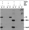
Exposure of the model substrates to increased temperature causes aggregation that is prevented by Hsp21. Proteins, 1 μM citrate synthase (CS) or 1 μM malate dehydrogenase (MDH) were incubated for 20 min at 47°C in the absence or presence of Hsp21 (12 μM). After centrifugation, soluble protein (supernatant, S) was separated from aggregated protein (pellet, P) and examined by SDS-PAGE; protein corresponding to 2.5, 1.8, and 12.5 μg loaded per lane of CS, MDH and Hsp21, respectively. The gel was silver stained.
Figure 3.
Figure 3.
Chemical cross-linking of the Hsp21 dodecamer with DTSSP. Hsp21 (12 μM) was pre-incubated at 25°C or 47°C for 20 min, after which the chemical cross-linker DTSSP (0.5 or 5 mM) was added for 20 min. Samples were lyophilized and dissolved in nonreducing SDS-PAGE loading buffer (5 μg/lane). (A) No cross-linker; (B) 0.5 or 5 mM cross-linker; (C) 0.5 mM cross-linker, Hsp21 oxidized with (10 mM H2O2, 20 min) before use; (D) same as in A, but 50 mM DTT added prior to SDS-PAGE. The gel was silver stained.
Figure 4.
Figure 4.
Chemical cross-linking between Hsp21 and CS with DTSSP. Hsp21 (12 μM) and CS (1 or 10 μM) were preincubated at 25°C or 47°C for 20 min, after which the chemical cross-linker DTSSP (5 mM) was added. (A) Nonreduced SDS-PAGE; (B) SDS-PAGE with 50 mM DTT added to samples prior to loading to reduce the disulphide bridge in the cross-linker. The gel was silver stained.
Figure 5.
Figure 5.
MALDI mass spectra used to identify cross-linked dipeptides. Mass spectra recorded for tryptic digests of Hsp21 control (top), Hsp21 with DTSSP (middle), and Hsp21 with DTSSP and DTT (bottom), showing how the peak (MH+ 1527.58), which corresponds to a cross-linked dipeptide, appears upon addition of the cross-linker DTTSP and disappears upon addition of DTT, which reduces the disulphur bond in the cross-linker. A new peak corresponding to a single peptide with half the cross-linker in alkylated form appears (MH+ 822.33) in the bottom spectra.
Figure 6.
Figure 6.
Multiple sequence alignment of Hsp21 and Hsp16.9 Secondary structure elements are marked below the sequences according to the structure determined to atomic resolution for Hsp16.9 (1GME). The four hydrophobic sites suggested to become exposed by dodecamer disassembly to dimers (van Montfort et al. 2001b) are color coded in the following way: The IXI/V-motif in the C-terminal tail, which in Hsp21 and its orthologs is extended to IXVXI, including three hydrophobic residues I179, V181, I183, (yellow); the C-terminal binding groove covered by the IXI/V-motif (red); the N-terminal arm (green); the patch in α-crystallin domain covered by the N-terminal arm (purple). Lysine residues are marked in blue. Sequence alignment was performed with Clustal W (http://www.ebi.ac.uk/clustalw/).
Figure 7.
Figure 7.
Hsp21 modeled onto the Hsp16.9 structure. (A) Top view of the dodecamer showing the double-disc structure with an upper and a lower ring of three dimers and six of 12 N-terminal arms forming intertwined α-helices in the center of the dodecamer. (B) Side view showing one of the three tetramers. In each tetramer the dimers are stabilized through strand exchange between monomers and dimer–dimer stabilization obtained by the C-terminal tail of one of the monomers (the one with disordered N terminus) in each dimer strapping across to the partner dimer in the other disc (encircled, corresponding to cross-linked dipeptides MH+ 2252 and 1506) (Table 2). (C) Side view showing the contact region between two adjacent tetramers formed by the N-terminal α-helices of one monomer (light blue) in a dimer in the upper disc and one monomer (pink) in a dimer in the lower disc (eclipsed, corresponding to cross-linked dipeptides MH+ 1527, 1785, 3209, and 2421) (Table 2). Stabilization of tetramers is also obtained by the C-terminal tail of one of the monomers (the one with an ordered N terminus) in each dimer strapping across to the partner dimer in the neighboring tetramer (encircled, corresponding to cross-linked dipeptides MH+ 2252, 1506) (Table 2). Close-up shows the C-terminal binding groove (red) in one monomer, formed by residues V109, I111, and V113 (β-strand 4), and I156, A158, and L160 (β-strand 8), covered by the C-terminal tail (yellow) with the three hydrophobic residues I179, V181, and I183, belonging to a neighbor tetramer (cyan). The Hsp21 model was generated using the PDB-file 1GME and the chains A and B used as the template structure for a homology model created by 3D-JIGSAW. (http://www.bmm.icnet.uk/∼3djigsaw/ and the program Chimera at http://www.cgl.ucsf.edu/chimera were used to generate images.)
Figure 8.
Figure 8.
The CS dimer and lysine residues involved in interaction with Hsp21. (A) All lysines in CS marked in blue. (B) All lysines detected in cross-linked CS-Hsp21 dipeptides marked in purple. (C) Lysine residues detected in cross-linked CS-Hsp21 dipeptides involving different suggested substrate-binding regions in Hsp21 color coded according to Figure 6 in the following way: the IXI/V-motif in the C-terminal tail (yellow), the C-terminal binding groove covered by the IXI/V-motif (red), and the N-terminal arm (green). The image was generated using the program Chimera (http://www.cgl.ucsf.edu/chimera) and the PDB-file 4CTS.
Figure 9.
Figure 9.
Increasing numbers of available sHsp substrate-binding sites. The number of substrate-binding sites can increase gradually with an increasing amount of substrate. (A) Low amount of substrate. In the structure determined for Hsp16.9 (van Montfort et al. 2001b), every second N-terminal arm is invisible, since it is not interacting with another N-terminal arm, and therefore not ordered. These six flexible arms, each containing two substrate-binding sites (color-coded green and purple in Fig. 6) may be available for interaction with substrates without dodecamer disassembly. (B) Larger amounts of substrate: Via dynamic subunit exchange, or dodecamer disassembly, more substrate-binding sites are mobilized. The number of available substrate-binding sites increases gradually, from 12 (in dodecamer) to 24 (if tetramers) to 36 (if dimers) to 72 (if monomers) as calculated below, incorporated into large sHsp–substrate complexes (for simplicity only tetramer-based complexes are visualized here). Numbers of substrate-binding sites hidden in the dodecamer in pairwise hydrophobic patch interactions: assuming that each monomer has six potential substrate-binding sites (the color-coded four sites in Fig. 6, plus the loop at the dimer interface indicated in substrate binding by the cross-linked Hsp21–CS dipeptides containing K125/126 [Table 3], plus the surface this loop interacts with in the dimer). In the dodecamer, 12 available substrate-binding sites are offered by the flexible N-terminal arms and their patches. If tetramers 24 (all 12 N-terminal arms and their patches); if dimers 36 (add 12, the six C-terminal tails that cover six patches, within tetramers and between tetramers); if monomers 72 (add 12, the C-terminal tails that cover six patches within dimers, plus 12 more, the released loops and their patches) substrate-binding sites become exposed.

References

    1. Åhrman E., Gustavsson, N., Hultschig, C., Boelens, W., and Sundby Emanuelsson, C. E. 2007. Small heat shock proteins prevent aggregation of citrate synthase and bind to the N-terminal region which is absent in thermostable forms of citrate synthase. Extremophiles (in press). - PubMed
    1. Aquilina J.A. and Watt, S.J. 2007. The N-terminal domain of αB-crystallin is protected from proteolysis by bound substrate. Biochem. Biophys. Res. Commun. 353: 1115–1120. - PubMed
    1. Aquilina J.A., Benesch, J.L., Bateman, O.A., Slingsby, C., and Robinson, C.V. 2003. Polydispersity of a mammalian chaperone: Mass spectrometry reveals the population of oligomers in αB-crystallin. Proc. Natl. Acad. Sci. 100: 10611–10616. - PMC - PubMed
    1. Arnott M.A., Michael, R.A., Thompson, C.R., Hough, D.W., and Danson, M.J. 2000. Thermostability and thermoactivity of citrate synthases from the thermophilic and hyperthermophilic archaea, Thermoplasma acidophilum and Pyrococcus furiosus . J. Mol. Biol. 304: 657–668. - PubMed
    1. Basha E., Lee, G.J., Breci, L.A., Hausrath, A.C., Buan, N.R., Giese, K.C., and Vierling, E. 2004. The identity of proteins associated with a small heat shock protein during heat stress in vivo indicates that these chaperones protect a wide range of cellular functions. J. Biol. Chem. 279: 7566–7575. - PubMed

Publication types

MeSH terms

LinkOut - more resources