. 2007 Jul;16(7):1329-37.
doi: 10.1110/ps.062689807. Epub 2007 Jun 13.

Solvent-induced differentiation of protein backbone hydrogen bonds in calmodulin

Affiliations

Solvent-induced differentiation of protein backbone hydrogen bonds in calmodulin

Nenad Juranić et al. Protein Sci. 2007 Jul.

Abstract

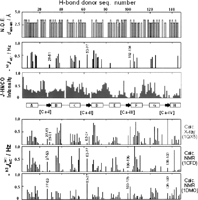
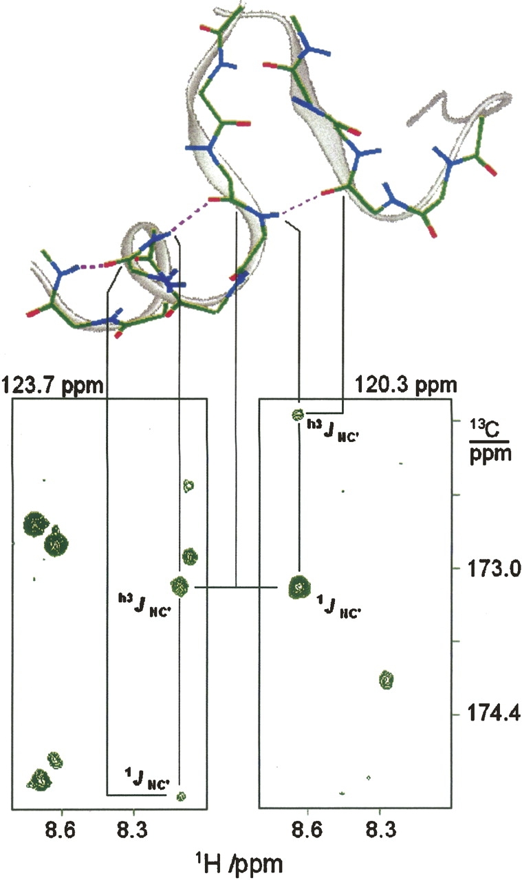
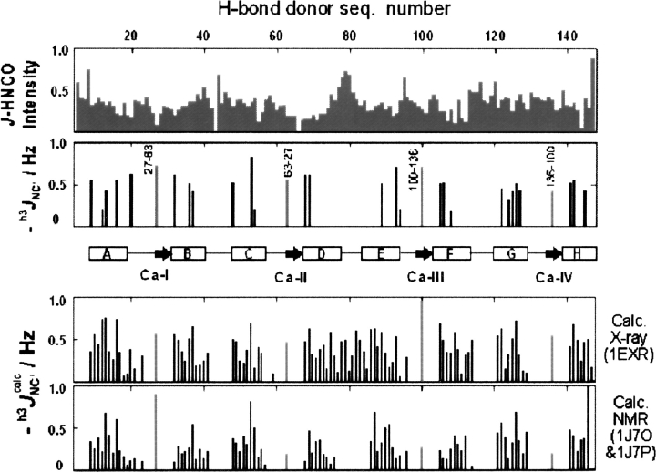
In apo and holoCaM, almost half of the hydrogen bonds (H-bonds) at the protein backbone expected from the corresponding NMR or X-ray structures were not detected by h3JNC' couplings. The paucity of the h3JNC' couplings was considered in terms of dynamic features of these structures. We examined a set of seven proteins and found that protein-backbone H-bonds form two groups according to the h3JNC' couplings measured in solution. H-bonds that have h3JNC' couplings above the threshold of 0.2 Hz show distance/angle correlation among the H-bond geometrical parameters, and appear to be supported by the backbone dynamics in solution. The other H-bonds have no such correlation; they populate the water-exposed and flexible regions of proteins, including many of the CaM helices. The observed differentiation in a dynamical behavior of backbone H-bonds in apo and holoCaM appears to be related to protein functions.


Figures

Figure 1.
Figure 1.
Sequential NOE distances of amide protons in apoCaM (Zhang et al. 1995a) (top, backbone h3 J NC′ couplings [second from top]) and relative sensitivity of J-HNCO experiment (third from top). (Central insert) The secondary structure of apoCaM; (rectangular boxes) helices; (arrows) the short β-sheet contacts. Detected β-sheet contacts are labeled with the corresponding residue numbers. (Three bottom inserts) Magnitudes of the h3 J NC′ calc couplings that we calculated for three of the reported structures (Kuboniwa et al. 1995; Zhang et al. 1995a) (in the 1QX5 structure [Schumacher et al. 2004], there are several intermolecular H-bonds [light-gray bars]).
Figure 2.
Figure 2.
Strips from 3D HNCO-J spectrum displaying NC′ correlation peaks in the H-bond chain 107/108 → 104/105 → 101/102 → 102/134. The spectrum, recorded at the J-coupling evolution time of 1/4J = 32 msec, is the sum of two 3D HNCO-J spectra each collected for 4 d on a Bruker 600 MHz spectrometer equipped with a cryo-probe.
Figure 3.
Figure 3.
Relative sensitivity of J-HNCO experiment (top) and backbone h3 J NC′ couplings (second from top). (Central insert) The secondary structure of holoCaM; (rectangular boxes) helices; (arrows) the short β-sheet contacts. Detected β-sheet contacts are labeled with the corresponding residue numbers. (Bottom inserts) The magnitudes of the h3 J NC′ calc couplings that we calculated for two of the reported structures (Wilson and Brunger 2000; Chou et al. 2001).
Figure 4.
Figure 4.
Hydrophobic contact of helices E and H in holoCaM. (Dashed lines) The H-bonds detected by h3 J NC′ couplings (magenta-colored region of the helices). (Top part) The peptide-bond 1 J NC′ couplings in the E-helix of apo and holoCAM.
Figure 5.
Figure 5.
Angle–distance correlation of H-bonds from seven proteins (crambin, ubiquitin, protein G, parvalbumin, IFABP, and apo- and holo-calmodulin) divided into two subsets according to the magnitude of the H-bond couplings (|h3 J NC′| > 0.2 Hz, top; |h3 J NC′| < 0.1 Hz, bottom). The distance correlation for the donor angle, ΔαH (○), is matched with the correlation for the acceptor angle, ΔαO (□), upon scaling the latter by 0.72. (The full curved line) The fifth-power distance dependence (0.8d OH 5); (dashed lines) ∼90% containment boundaries. The peptide groups involved in the coordination to Ca2+ were excluded from the analysis.
Figure 6.
Figure 6.
Experimental RDCs of peptide groups (Δ1 J HN, Δ1 J NC/0.12, and Δ1 J CCa/0.20) in α-helices of the holoCaM C terminus, in comparison with the calculated values for the corresponding crystal and solution structures. The crystal structure is represented by X-ray high-resolution structure 1exr-model-A (Wilson and Brunger 2000), while the solution structure is modeled from the X-ray structure to satisfy RDCs. The high-J and low-J data refer to the size of proton-donating H-bonds of the peptide groups. (Lower insert) The X-ray and the modeled solution structure of helix E. (Small arrows) Connection between peptide-group orientations and RDC fit for the peptide group in position 91/92. The hydrophobic anchoring residue is indicated.
Figure 7.
Figure 7.
Comparison of the high-J subset H-bonds in apoCaM (top) and holoCaM (bottom). The population of the helices with the detected H-bonds is represented by the shades of the rectangular boxes: (black) >70%; (gray) ∼50%; (empty) <30%. Detected β-sheet contacts are labeled with the corresponding residue numbers.

Comment in

Similar articles

Cited by

References

    1. Alexandrescu A.T., Snyder, D.R., and Abildgaard, F. 2001. NMR of hydrogen bonding in cold-shock protein A and an analysis of the influence of crystallographic resolution on comparisons of hydrogen bond lengths. Protein Sci. 10: 1856–1868. - PMC - PubMed
    1. Assadi-Porter F.M., Abildgaard, F., Blad, H., and Markley, J.L. 2003. Correlation of the sweetness of variants of the protein brazzein with patterns of hydrogen bonds detected by NMR spectroscopy. J. Biol. Chem. 278: 31331–31339. - PubMed
    1. Barabato G., Ikura, M., Kay, L.E., Pastor, R.W., and Bax, A. 1992. Backbone dynamics of calmodulin studied by 15N relaxation using inverse detected two-dimensional NMR spectroscopy: The central helix is flexible. Biochemistry 31: 5269–5278. - PubMed
    1. Barfield M. 2002. Structural dependencies of interresidue scalar coupling h3 J NC, and donor 1H chemical shifts in the hydrogen bonding regions of proteins. J. Am. Chem. Soc. 124: 4158–4168. - PubMed
    1. Bouvignies G., Bernado, P., Meier, S., Cho, K., Grzesiek, S., Bruschweiler, R., and Blackledge, M. 2005. Identification of slow correlated motions in proteins using residual dipolar and hydrogen-bond scalar couplings. Proc. Natl. Acad. Sci. 102: 13885–13890. - PMC - PubMed

LinkOut - more resources