. 2007 Jun 19;104(25):10595-600.
doi: 10.1073/pnas.0702072104. Epub 2007 Jun 13.

IGF binding protein-3 regulates hematopoietic stem cell and endothelial precursor cell function during vascular development

Affiliations

IGF binding protein-3 regulates hematopoietic stem cell and endothelial precursor cell function during vascular development

Kyung-Hee Chang et al. Proc Natl Acad Sci U S A. .

Abstract

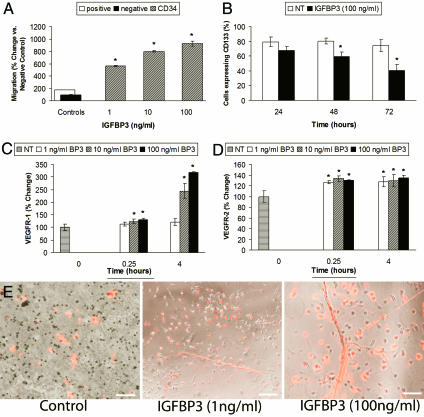
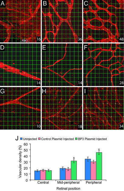
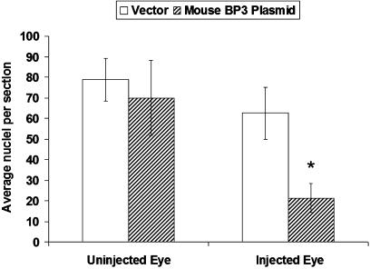
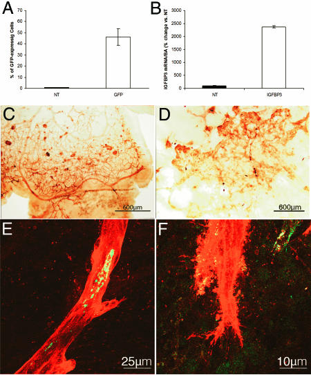
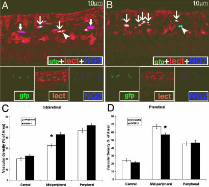
We asked whether the hypoxia-regulated factor, insulin-like growth factor binding protein-3 (IGFBP3), could modulate stem cell factor receptor (c-kit+), stem cell antigen-1 (sca-1+), hematopoietic stem cell (HSC), or CD34+ endothelial precursor cell (EPC) function. Exposure of CD34+ EPCs to IGFBP3 resulted in rapid differentiation into endothelial cells and dose-dependent increases in cell migration and capillary tube formation. IGFBP3-expressing plasmid was injected into the vitreous of neonatal mice undergoing the oxygen-induced retinopathy (OIR) model. In separate studies, GFP-expressing HSCs were transfected with IGFBP3 plasmid and injected into the vitreous of OIR mice. Administering either IGFBP3 plasmid alone or HSCs transfected with the plasmid resulted in a similar reduction in areas of vasoobliteration, protection of the developing vasculature from hyperoxia-induced regression, and reduction in preretinal neovascularization compared to control plasmid or HSCs transfected with control plasmid. In conclusion, IGFBP3 mediates EPC migration, differentiation, and capillary formation in vitro. Targeted expression of IGFBP3 protects the vasculature from damage and promotes proper vascular repair after hyperoxic insult in the OIR model. IGFBP3 expression may represent a physiological adaptation to ischemia and potentially a therapeutic target for treatment of ischemic conditions.


Conflict of interest statement

The authors declare no conflict of interest.

Figures

Fig. 1.
Fig. 1.
IGFBP3 modulates CD34+ cell behavior in vitro. (A) CD34+ cells (hatched bars) migrated in a dose-dependent manner toward IGFBP3. Migration is measured by relative fluorescent units (RFUs) compared to negative control. Negative control, medium alone arbitrarily set at 100 (filled bar); positive control, HPGM medium containing 20% serum (open bar). (B) CD34+ cells exposed to IGFBP3 (filled bars) for 48 and 72 h demonstrate reduced CD133 expression, supporting that IGFBP3 promotes their differentiation toward endothelial cells (P < 0.05 vs. control shown in open bars). (C) IGFBP3 exposure significantly increased VEGFR1 expression in CD34+ cells at the higher concentrations tested. ∗, P < 0.05 vs. control for 15 min and P < 0.001 for 4 h. (D) IGFBP3 increased the expression of VEGFR2 by 26.58% (∗, P < 0.001) at 15 min of exposure and by 27.5% (P < 0.001) at 4 h of exposure. (E) IGFBP3 increases EPC proliferation and tube formation compared to cells treated with control medium. IGFBP3 exposure resulted in a dose-dependent increase in tube formation. The cells have been exposed to fluorescently labeled acetylated LDL. The labeled cells appearing “red” in color represent EPCs that have differentiated into endothelial cells. (Magnification: ×100.) (Scale bars: Left and Center, 150 μm; Right, 100 μm.)
Fig. 2.
Fig. 2.
Quantitative measure of vascular density in the OIR model. Representative fields of view from each of the central, midperipheral, and peripheral retinas captured by using the ×40 objective lens. A 10 × 10 grid was superimposed onto the micrograph, and the incidence of presence of vessels at the intersection points of each grid was determined. The measurement of vascular density was expressed as a percentage from 0 to 100 (bottom right corner of the image). Fields of view selected for analysis included regions of capillary-sized vessels directly adjacent to radial arterioles. (A–C) Images taken from eyes injected with IGFBP3 plasmid. Pups were placed in hyperoxia for 5 days and then normoxia for 5 days. (D–F) Images taken from contralateral uninjected eyes from the OIR group. (G–I) Images taken from empty plasmid-injected eyes from the OIR group. (J) One-way ANOVA showed that IGFBP3 protects the retinal vasculature from hyperoxia-induced vessel regression in midperipheral [F(2, 42) = 36.40; ∗, P < 0.001] and peripheral [F(2, 42) = 32.33; ∗, P < 0.001] regions, but did not have any significant effect on the central region of the retina [F(2, 42) = 0.37, P > 0.05].
Fig. 3.
Fig. 3.
Intravitreal injection with IGFBP3-expressing plasmid reduces preretinal neovascularization. Neovascularization was evaluated by measuring the reduction in preretinal endothelial nuclei in pups injected with IGFBP3-expressing plasmid (hatched bars) in one eye compared with control pups that received empty plasmid in one eye (n = 9, open bars). Uninjected eyes were from the same pups. ∗, P < 0.005 when comparing control plasmid-injected eyes to IGFBP3 plasmid-injected eyes.
Fig. 4.
Fig. 4.
Retinas from eyes injected intravitreally with HSCs transfected with IGFBP3 and underwent the OIR model. (A) HSCs were transfected with 10 μg of DNA of a GFP plasmid (open bars) by using either Lipofectamine transfection reagent or polyethyleneimine. Mock-transfected cells did not contain any DNA (filled bars). GFP expression in the cells was assessed by flow-cytometry analysis. Cells that underwent mock transfection did not show GFP cells, whereas polyethyleneimine showed increased transfection compared to Lipofectamine. (B) Transfection of GFP HSC with IGFBP3-expressing plasmid (open bars) results in a 25-fold increase in IGFBP3 expression in vitro compared with nontransfected (NT) controls (filled bars). (C and D) Low-magnification views of GS isolectin-labeled vasculature in the retina from a pup injected with IGFBP3-transfected HSCs (C) compared with the contralateral uninjected eye (D). (C, E, and F) HSC-IGFBP3 animal showed a vascular tree with more normal morphology, with a mature pattern of differentiation, normal dichotomous branching pattern, and less abnormal vascularization. (E and F) Retinal flat mounts demonstrate the localization of GFP HSCs within the vasculature. Merged green (HSC) and red (resident vasculature) channels demonstrating the localization of HSCs within the GS isolectin-labeled vasculature (red). There is evidence of GFP HSC incorporation (yellow) in vascular endothelial cells lining neovascular lumens (E) and filopodia spread from neovascular clumps (F).
Fig. 5.
Fig. 5.
IGFBP3-expressing HSCs give rise to vascular endothelial and perivascular cells, and injection of IGFBP3 HSCs during the hypoxic phase of the OIR model results in decreased preretinal neovascularization. (A and B) Cryostat sections from IGFBP3-injected eyes showing GFP HSC-derived cells giving rise to vascular endothelial as well as perivascular cells. Isolectin stains endothelial and perivascular cells, whereas NG2 is a pericyte marker. Thin arrows in A and B mark triple-labeled (NG2+/GFP+/isolectin+) cells representing vasculature where GFP HSCs have become endothelial cells or pericytes. Thick arrows (A) show two NG2+/isolectin/GFP+ HSC-derived pericytes within regions of the resident vasculature. Arrowhead in A shows an NG2/isolectin+/GFP+ HSC-derived endothelial cell (yellow). Arrowhead in B shows an NG2+/isolectin/GFP+ resident pericyte (pale blue). (C and D) Vascular density measured from the intraretinal vessels (C) or from the preretinal vessels (D). (C) Vascular density measurements (intraretinal vessels) showed a significant difference in the vessels in the midperipheral region between eyes injected with HSC transfected with control plasmid vs. HSCs transfected with IGFPB3 plasmid (∗, P < 0.003). (D) Vascular density measurements (preretinal vessels) showed a significant difference in the vessels in the midperipheral region between eyes injected with HSC transfected with control plasmid vs. HSCs transfected with IGFBP3 plasmid (∗, P < 0.0008).

References

    1. Smith LE, Shen W, Perruzzi C, Soker S, Kinose F, Xu X, Robinson G, Driver S, Bischoff J, Zhang B, et al. Nat Med. 1999;5:1390–1395. - PubMed
    1. Hellstrom A, Engstrom E, Hard AL, Albertsson-Wikland K, Carlsson B, Niklasson A, Lofqvist C, Svensson E, Holm S, Ewald U, et al. Pediatrics. 2003;112:1016–1020. - PubMed
    1. Lofqvist C, Engstrom E, Sigurdsson J, Hard AL, Niklasson A, Ewald U, Holmstrom G, Smith LE, Hellstrom A. Pediatrics. 2006;117:1930–1938. - PubMed
    1. Grant MB, Russell B, Fitzgerald C, Merimee TJ. Diabetes. 1986;35:416–420. - PubMed
    1. Grant MB, Wargovich TJ, Ellis EA, Tarnuzzer R, Caballero S, Estes K, Rossing M, Spoerri PE, Pepine C. Regul Pept. 1996;67:137–144. - PubMed

Publication types

MeSH terms

Substances