Clinical Trial
. 2007 Jun 19;104(25):10589-94.
doi: 10.1073/pnas.0702031104. Epub 2007 Jun 13.

IGFBP3 suppresses retinopathy through suppression of oxygen-induced vessel loss and promotion of vascular regrowth

Affiliations
Clinical Trial

IGFBP3 suppresses retinopathy through suppression of oxygen-induced vessel loss and promotion of vascular regrowth

Chatarina Lofqvist et al. Proc Natl Acad Sci U S A. .

Abstract

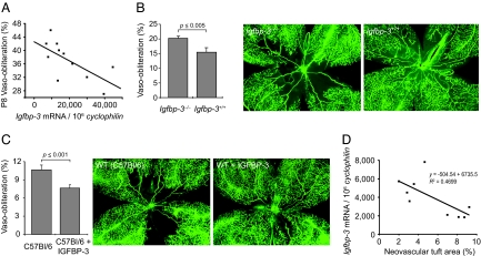
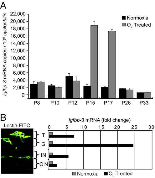
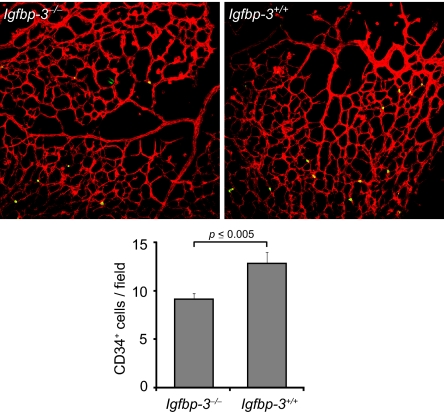
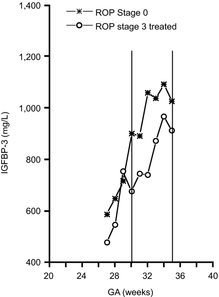
Vessel loss precipitates many diseases. In particular, vessel loss resulting in hypoxia induces retinal neovascularization in diabetic retinopathy and in retinopathy of prematurity (ROP), major causes of blindness. Here we define insulin-like growth factor binding protein-3 (IGFBP3) as a new modulator of vascular survival and regrowth in oxygen-induced retinopathy. In IGFBP3-deficient mice, there was a dose-dependent increase in oxygen-induced retinal vessel loss. Subsequent to oxygen-induced retinal vessel loss, Igfbp3(-/-) mice had a 31% decrease in retinal vessel regrowth versus controls after returning to room air. No difference in serum insulin-like growth factor 1 (IGF1) levels was observed among groups. Wild-type mice treated with exogenous IGFBP3 had a significant increase in vessel regrowth. This correlated with a 30% increase in endothelial progenitor cells in the retina at postnatal day 15, indicating that IGFBP3 could be serving as a progenitor cell chemoattractant. In a prospective clinical study, we measured IGFBP3 (and IGF1) plasma levels weekly and examined retinas in all premature infants born at gestational ages <32 weeks at high risk for ROP. The mean level of IGFBP3 at 30-35 weeks postmenstrual age (PMA) for infants with proliferative ROP (ROP stages 3>, n = 13) was 802 microg/liter, and for infants with no ROP (ROP stage 0, n = 38) the mean level was 974 microg/liter (P < 0.03). These results suggest that IGFBP3, acting independently of IGF1, helps to prevent oxygen-induced vessel loss and to promote vascular regrowth after vascular destruction in vivo in a dose-dependent manner, resulting in less retinal neovascularization.


Conflict of interest statement

The authors declare no conflict of interest.

Figures

Fig. 1.
Fig. 1.
IGFBP3 protects against oxygen-induced vessel loss. (A) Igfbp3 mRNA expression is associated with protection against oxygen-induced retinal vessel loss in a dose-dependent manner at P8 after 18 h of 75% oxygen in Igfbp3+/− mice (n = 11 mice, with mean of two retinas at each point) (r2 = −0.70; P ≤ 0.006). (B) (Center and Right) Loss of IGFBP3 decreases retinal vessel regrowth after oxygen-induced loss (P17). IGFBP3 protection after oxygen-induced vessel loss persists as shown at P17 in wild-type (n = 38 eyes) (Right) compared to Igfbp3−/− mice (n = 52 eyes) (Center). (Left) Fractional retinal area of vasoobliteration in Igfbp3−/− mice was 20.2 ± 0.1% (SEM) compared to wild-type mice with an area of 15.1 ± 0.5% (P ≤ 0.005) (Right). Representative retinal whole mounts show less vessel loss in wild-type than in Igfbp3−/− mice. (C) Exogenous IGFBP3 further increases vessel regrowth over baseline. (Center and Right) Whole-mounted retinas of C57BL/6 mice treated with saline after oxygen-induced vessel loss (Center) have decreased vessel regrowth centrally than littermates treated with i.p. IGFBP3 (Right) from P12 to P14. (Left) There is a 40% decrease in retinal area without vessels with IGFBP3 treatment compared to control (P ≤ 0.001) as seen in whole-mounted retinas. (D) Retinal NV decreases with increasing IGFBP3. Igfbp3 mRNA expression is associated with protection in a dose-dependent manner against retinal NV in the ROP mouse model at P17 in Igfbp3+/− and Igfbp3+/+ mice (n = 11 mice, with mean of two retinas at each point) (r2 = −0.47).
Fig. 2.
Fig. 2.
Igfbp3 mRNA in whole retina increases with hypoxia at P15 and P17 in whole retina. (A) Each bar represents Igfbp3 mRNA copy number normalized to 1 million copies of cyclophilin (an unchanging control gene). During hypoxia at P15 and P17 in O2-treated mice, Igfbp3 expression increased ≈9-fold over normoxic control (n = 12 for each group). (B) Igfbp3 mRNA is associated with retinal blood vessels and increases with hypoxia. Quantitative real-time RT-PCR analysis of Igfbp3 mRNA analyzed from laser-captured areas of retinal blood vessels shows a large increase in Igfbp3 mRNA in the superficial vascular layers at P17 with hypoxia, indicating that IGFBP3 is associated with retinal blood vessels and not with surrounding tissue. T, neovascular vessel tufts extending into the vitreous; G, ganglion cell layer; ON, outer nuclear or photoreceptor layer; IN, inner nuclear layer.
Fig. 3.
Fig. 3.
Decreased number of EPCs in the retina of IGFBP3-null mice. Quantification of CD34+ cells in the retina of Igfbp3−/− mice (Upper Left) at P15 after OIR (n = 8) revealed a significantly decreased number of EPCs (Lower) compared to wild-type animal (n = 7; P = 0.003) (Upper Right). Red, lectin-Alexa 594 (endothelial cell-specific); green, CD34-FITC antibody.
Fig. 4.
Fig. 4.
Lower serum levels of IGFBP3 are associated with reduction in ROP in children. The mean ± SEM level of IGFBP3 at 30–35 weeks PMA for infants with proliferative ROP was 802 ± 66 μg/liter and for infants with no ROP was 974 ± 41 μg/liter. The Mann–Whitney U test yielded a P value of 0.03, indicating a significant difference between the two groups in mean IGFBP3 at this time point.

References

    1. Hellstrom A, Engstrom E, Hard AL, Albertsson-Wikland K, Carlsson B, Niklasson A, Lofqvist C, Svensson E, Holm S, Ewald U, et al. Pediatrics. 2003;112:1016–1020. - PubMed
    1. Mohan S, Baylink DJ. J Endocrinol. 2002;175:19–31. - PubMed
    1. Firth SM, Baxter RC. Endocr Rev. 2002;23:824–854. - PubMed
    1. De Mellow JS, Baxter RC. Biochem Biophys Res Commun. 1988;156:199–204. - PubMed
    1. Conover CA. Endocrinology. 1992;130:3191–3319. - PubMed

Publication types

MeSH terms

Substances

LinkOut - more resources