. 2007 Jun 13;27(24):6363-73.
doi: 10.1523/JNEUROSCI.0307-07.2007.

Differential role of N-type calcium channel splice isoforms in pain

Affiliations

Differential role of N-type calcium channel splice isoforms in pain

Christophe Altier et al. J Neurosci. .

Abstract

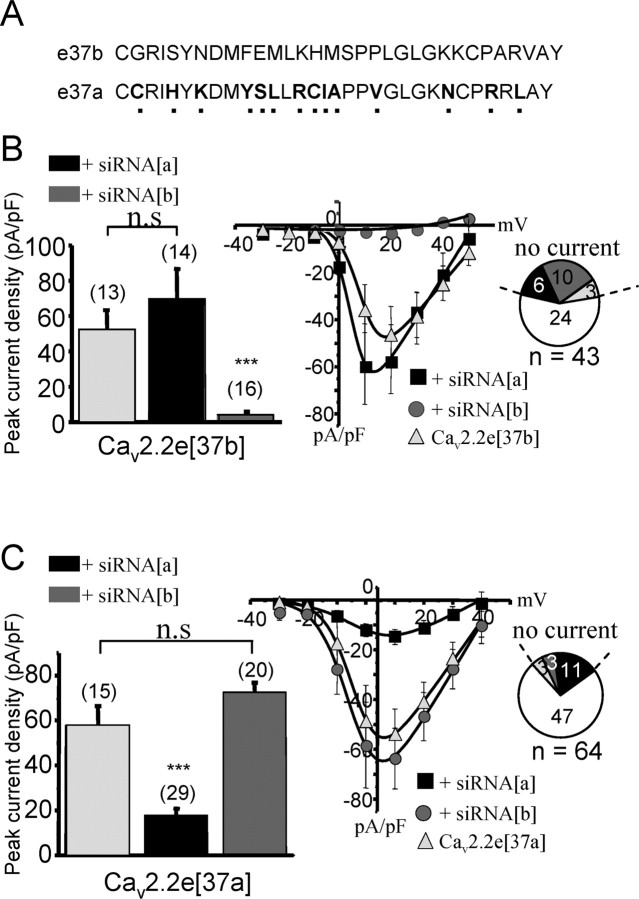
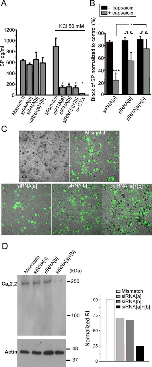
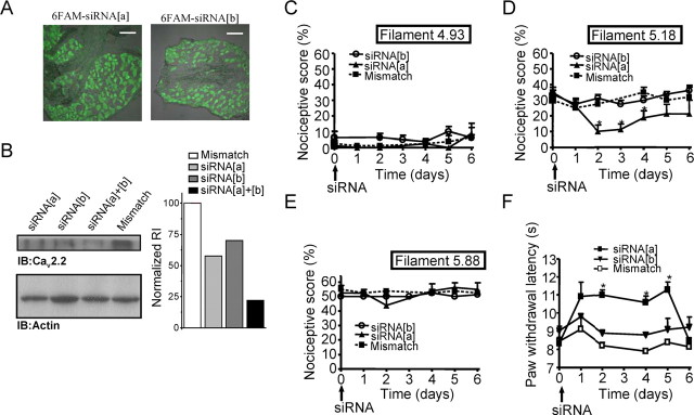
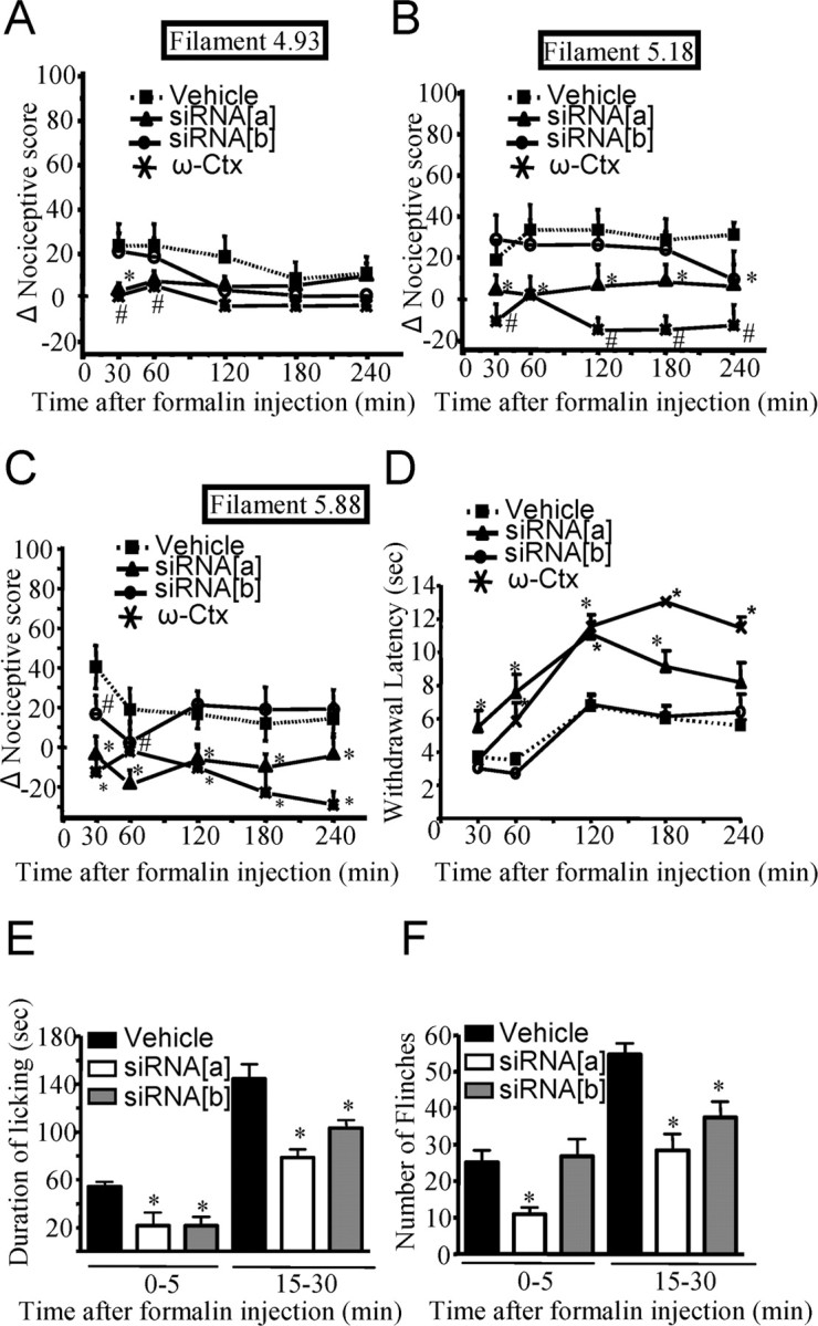
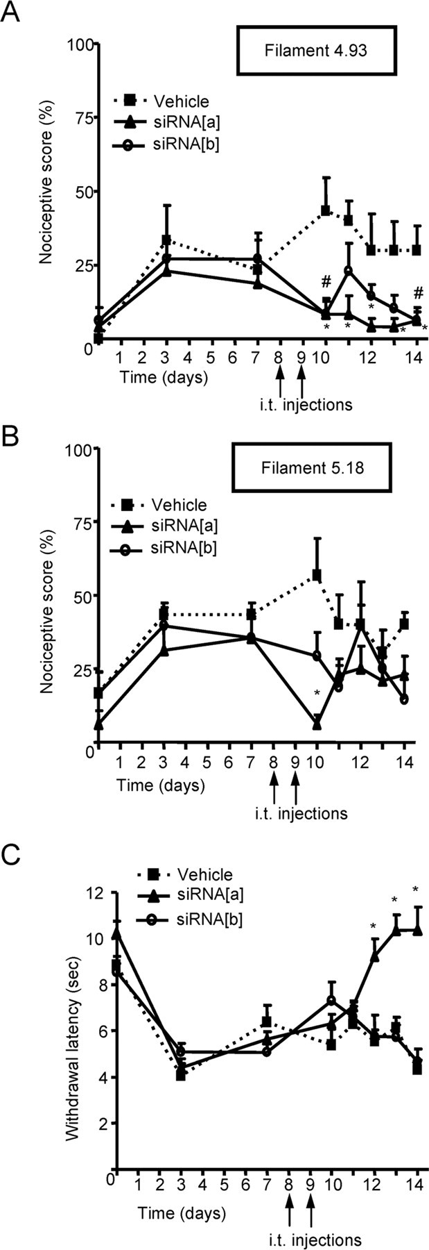
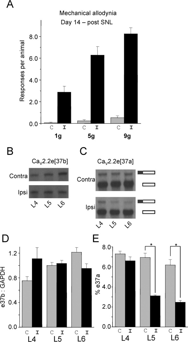
N-type calcium channels are essential mediators of spinal nociceptive transmission. The core subunit of the N-type channel is encoded by a single gene, and multiple N-type channel isoforms can be generated by alternate splicing. In particular, cell-specific inclusion of an alternatively spliced exon 37a generates a novel form of the N-type channel that is highly enriched in nociceptive neurons and, as we show here, downregulated in a neuropathic pain model. Splice isoform-specific small interfering RNA silencing in vivo reveals that channels containing exon 37a are specifically required for mediating basal thermal nociception and for developing thermal and mechanical hyperalgesia during inflammatory and neuropathic pain. In contrast, both N-type channel isoforms (e37a- and e37b-containing) contribute to tactile neuropathic allodynia. Hence, exon 37a acts as a molecular switch that tailors the channels toward specific roles in pain.


Figures

Figure 1.
Figure 1.
Electrophysiological evidence of the specificity of siRNA silencing. A, Sequence alignment between exons 37a and 37b of the rat CaV2.2 calcium channel α1 subunit. B, Whole-cell recordings of peak current densities of tsA-201 cells transfected with the CaV2.2e[37b] (+ β1b and α2–δ) isoform and treated with different types of siRNA (left). Numbers in parentheses are numbers of cells patched, and the asterisks denote statistical significance between untreated, siRNA[a]-treated, and siRNA-treated cells. Right, Portion of cells with or without current for different siRNA treatments. Middle, Current–voltage relationships of CaV2.2e[37b]-transfected cells represented in the left and treated with siRNA[a] or siRNA [b]. C, Same as in B but for CaV2.2e[37a] isoform (+β1b and α2–δ). Note that, in this case, only siRNA[a] decreased CaV2.2e[37a] peak current density and amplitude of currents. The solid lines in the current–voltage relationships were obtained via the Boltzmann equation, and error bars denote SEs.
Figure 2.
Figure 2.
Effects of siRNA treatment on substance P release and CaV2.2 protein expression in cultured DRG neurons. A, Quantification of substance P release by ELISA. DRG neurons were plated at the same density and treated with 6-FAM labeled siRNA constructs (i.e., mismatch, siRNA[a], or siRNA[b]). The amount of substance P secreted by the DRG culture was measured by sampling either the culture medium (reflecting tonic release) or immediately after replacement with medium and subsequent depolarization with 50 mm KCl (reflecting evoked release). ω-Conotoxin GVIA at 1 μm was applied to the cultures to block N-type channels as indicated. In this case, the KCl solution was also supplemented with toxin. B, Inhibition of substance P release normalized to control with or without previous treatment of the neurons with (10 μm) capsaicin to deplete capsaicin-sensitive nociceptors of their neuropeptide contents. *p < 0.05, among the capsaicin-treated groups, significant difference between siRNA[a]- and siRNA[a + b]-injected animals. ***p < 0.01, significant difference among siRNA[a] groups, between capsaicin-treated and untreated DRG. C, Uptake of 6-FAM-tagged siRNA into dorsal root ganglia neurons 2 d after siRNA transfection. D, Western blot analysis of CaV2.2 channel protein in lysate of the DRG neurons used in A. Protein (15 μg) was loaded in every lane. The Western blot was probed with an anti- CaV2.2 calcium channel antibody. As a control, an actin antibody was used on a second gel to verify the amount of protein. The bar chart (right) is a quantification of relative integrated density value from each band. Data from three experiments are included in the bar chart; no statistical analysis was conducted. n.s., Not significant.
Figure 3.
Figure 3.
Effects of CaV2.2 splice isoform-specific siRNA silencing on acute nociception. A, Uptake and distribution of 6-FAM-tagged siRNA into dorsal root ganglia after siRNA injection. Three days after 6-FAM–siRNA intrathecal injection, DRG were harvested, and fluorescence of siRNA[a] and siRNA[b] was visualized in DRG slices. Scale bars, 0.2 mm. B, Western blot analysis of CaV2.2 channel protein from DRG lysate (L4–L6) obtained from rats treated with siRNA. The Western blot was probed with an anti-CaV2.2 calcium channel antibody. An antibody against actin was used on a second gel to verify the amount of protein. The bar chart (right) is a quantification of the relative integrated density value from each band. Collected DRG lysate samples were run blind. Data from three experiments are included in the bar chart; no statistical analysis was conducted. C–F, Kinetics of basal nociceptive responses to mechanical stimuli (von Frey filament 4.93, 4 g bending force; von Frey filament 5.18, 15 g bending force; and filament 5.88, 60 g bending force) as a function of time after siRNA treatment (C–E) or thermal stimulus (F) after intrathecal injection of siRNA. Data are expressed as mean ± SE; data are from seven rats per group. The asterisks and number symbols indicate significant difference from basal conditions (p < 0.05). IB, Immunoblot.
Figure 4.
Figure 4.
Effect of CaV2.2 splice isoform-specific siRNA silencing on inflammatory allodynia and hyperalgesia induced by plantar injection of Formalin. A–C, Change in nociceptive scores compared with basal measurements in response to a mechanical innocuous stimulus (4.93 von Frey filament of 4 g bending force; A) as a function of time after Formalin injection. Animals were injected with siRNA 2 d earlier. Noxious stimuli (5.18 and 5.88 von Frey filaments of 15 and 60 g bending force, respectively) (B, C) in rats injected intrathecally with siRNA[a], siRNA[b], or vehicle 2 d before the induction of inflammation by intraplantar injection of Formalin. D, Kinetics of the withdrawal latency in response to a thermal stimulus plotted against the time after intraplantar injection of Formalin in rats treated intrathecally with siRNA[a], siRNA[b], or vehicle 2 d before the experiment. E, F, Total time spent during licking (E) and number of flinches (F) for the periods of 0–5 or 15–30 min after Formalin injection in groups of rats injected with siRNA. Data are expressed as mean ± SE; data are from eight rats per group. Asterisks and number symbols denote significant difference from vehicle-treated group (p < 0.05). ω-Cx, ω-Conotoxin.
Figure 5.
Figure 5.
Effect of CaV2.2 splice isoform-specific siRNA silencing on neuropathic allodynia and hyperalgesia induced by sciatic nerve ligature. A–C, Kinetics of nociceptive scores (A, B) and paw-withdrawal latency (C) in response to either a mechanical innocuous stimulus (4.93 von Frey filament of 4 g bending force; A), a noxious stimulus (5.18 von Frey filament of 15 g bending force; B), or a thermal stimulus (C) in rats injected intrathecally with siRNA[a], siRNA[b] or vehicle, 8 and 9 d after the induction of sciatic nerve constriction injury. Data are expressed as mean ± SE; data are from eight rats per group. Asterisks and number symbols denote significant difference from vehicle-treated group (p < 0.05).
Figure 6.
Figure 6.
Regulation of CaV2.2e[37a] and CaV2.2e[37b] during neuropathy. A, Mechanical allodynia was evaluated by measuring nociceptive responses using different von Frey filaments 14 d after SNL in the ipsilateral (I; black) and contralateral (C; gray) side. B, RPA analysis using the e37b probe. We pooled total RNA isolated from DRG neurons from the same location (L4, L5, or L6) from animals presenting allodynia (n = 17). C, RPA analysis using the e37a probe. Total RNA is the same as in B. The top band in both contralateral and ipsilateral gels shows “fully protected” e37a probe and indicates the amount of CaV2.2e[37a] mRNA. The bottom band shows “partially protected” e37a probe and indicates non-e37a-containing mRNA (essentially CaV2.2e[37b] mRNA). D, Quantification of e37b mRNA protection levels presented as a ratio of e37b to GAPDH. E, Quantification of e37a mRNA protection levels represented as a percentage of total CaV2.2 mRNA (for calculation, see Materials and Methods). All data reflect the mean ± SE for three independent hybridizations of probe or probe pairs with the pooled DRG RNA.

Similar articles

Cited by

References

    1. Altier C, Zamponi GW. Targeting Ca2+ channels to treat pain: T-type versus N-type. Trends Pharmacol Sci. 2004;25:465–470. - PubMed
    1. Altier C, Khosravani H, Evans RM, Hameed S, Peloquin JB, Vartian BA, Chen L, Beedle AM, Ferguson SS, Mezghrani A, Dubel SJ, Bourinet E, McRory JE, Zamponi GW. ORL1 receptor-mediated internalization of N-type calcium channels. Nat Neurosci. 2006;9:31–40. - PubMed
    1. Beedle AM, McRory JE, Poirot O, Doering CJ, Altier C, Barrere C, Hamid J, Nargeot J, Bourinet E, Zamponi GW. Agonist-independent modulation of N-type calcium channels by ORL1 receptors. Nat Neurosci. 2004;7:118–125. - PubMed
    1. Bell TJ, Thaler C, Castiglioni AJ, Helton TD, Lipscombe D. Cell-specific alternative splicing increases calcium channel current density in the pain pathway. Neuron. 2004;41:127–138. - PubMed
    1. Bennett GJ, Xie YK. A peripheral mononeuropathy in rat that produces disorders of pain sensation like those seen in man. Pain. 1988;33:87–107. - PubMed

Publication types

MeSH terms

LinkOut - more resources