. 2007 Jun 13;27(24):6417-27.
doi: 10.1523/JNEUROSCI.0690-07.2007.

Brain-derived neurotrophic factor regulates cholesterol metabolism for synapse development

Affiliations

Brain-derived neurotrophic factor regulates cholesterol metabolism for synapse development

Shingo Suzuki et al. J Neurosci. .

Abstract

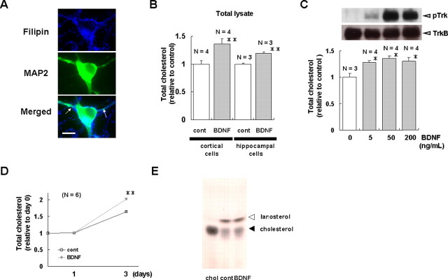
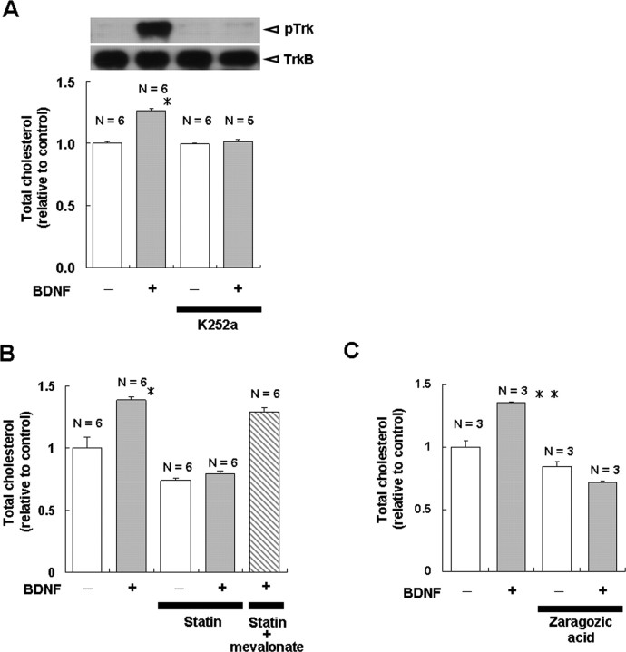
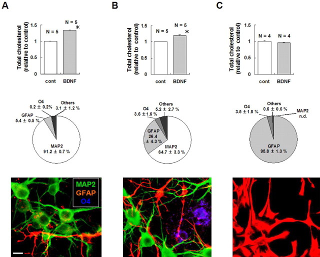
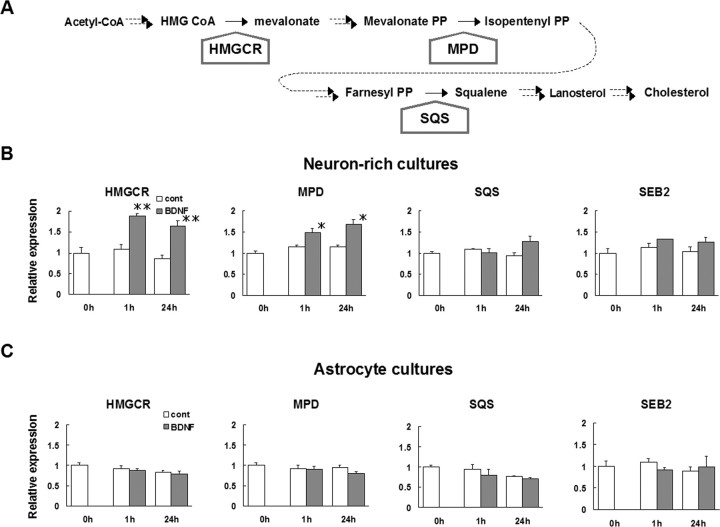
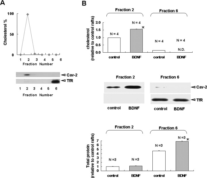
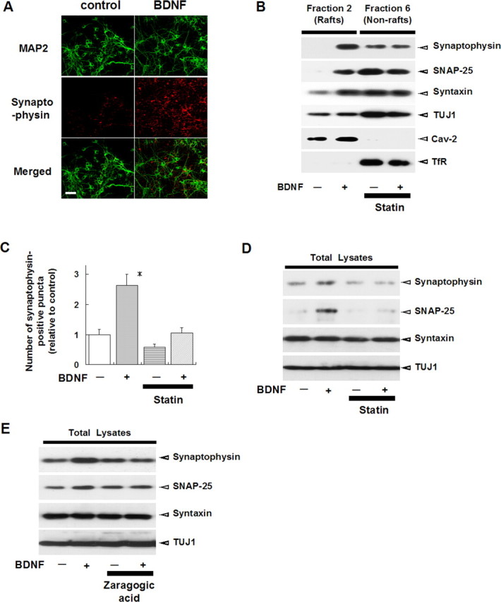
Brain-derived neurotrophic factor (BDNF) exerts multiple biological functions in the CNS. Although BDNF can control transcription and protein synthesis, it still remains open to question whether BDNF regulates lipid biosynthesis. Here we show that BDNF elicits cholesterol biosynthesis in cultured cortical and hippocampal neurons. Importantly, BDNF elicited cholesterol synthesis in neurons, but not in glial cells. Quantitative reverse transcriptase-PCR revealed that BDNF stimulated the transcription of enzymes in the cholesterol biosynthetic pathway. BDNF-induced cholesterol increases were blocked by specific inhibitors of cholesterol synthesis, mevastatin and zaragozic acid, suggesting that BDNF stimulates de novo synthesis of cholesterol rather than the incorporation of extracellular cholesterol. Because cholesterol is a major component of lipid rafts, we investigated whether BDNF would increase the cholesterol content in lipid rafts or nonraft membrane domains. Interestingly, the BDNF-mediated increase in cholesterol occurred in rafts, but not in nonrafts, suggesting that BDNF promotes the development of neuronal lipid rafts. Consistent with this notion, BDNF raised the level of the lipid raft marker protein caveolin-2 in rafts. Remarkably, BDNF increased the levels of presynaptic proteins in lipid rafts, but not in nonrafts. An electrophysiological study revealed that BDNF-dependent cholesterol biosynthesis plays an important role for the development of a readily releasable pool of synaptic vesicles. Together, these results suggest a novel role for BDNF in cholesterol metabolism and synapse development.


Figures

Figure 1.
Figure 1.
BDNF elicits cholesterol biosynthesis in cultured cortical and hippocampal neurons. Cholesterol content of cortical and hippocampal cells cultured under various conditions was measured. A, Cholesterol in cultured cortical neurons was shown by staining cells with filipin, as described previously (Ma et al., 2003). Scale bar, 10 μm. B, Cortical and hippocampal cells were cultured in the presence or absence of 200 ng/ml BDNF for 3 and 5 d, respectively. Compared with untreated cells (cont), BDNF-treated cortical and hippocampal cells had higher cholesterol contents. C, BDNF increased the cholesterol content in cortical cells in a dose-dependent manner. Top, The TrkB receptor was activated by BDNF in a dose-dependent manner. Cell lysates were collected before or 3 min after treatment with the indicated concentration of BDNF. Immunoblotting was performed with anti-TrkB and anti-phospho-Trk antibodies, as described previously (Suzuki et al., 2004). Bottom, BDNF increased cholesterol content in a concentration-dependent manner. Cholesterol content was measured 3 d after treatment with the indicated concentration of BDNF. D, A time course study of the BDNF-dependent cholesterol increase in cortical cells. Cells were cultured in the presence or absence of BDNF (200 ng/ml) for the indicated times, and the cholesterol amount was determined. E, TLC analysis of cholesterol in BDNF-treated cortical neurons. Sterols were isolated from cortical neurons cultured in the presence or absence of BDNF (200 ng/ml) for 5 d. Extracts were dissolved in isopropyl alcohol and separated by TLC. Spots were visualized by using p-anisaldehyde. The positions of cholesterol and lanosterol are indicated with arrowheads. As a control, commercially obtained, purified cholesterol (chol) was loaded. Densitometric analysis demonstrated that the increase in cholesterol content in response to BDNF was 38 ± 12%. The value for each cholesterol band was normalized to that of the control sample. This and all other figures demonstrate that results are relative to control and are shown as the means ± SEM. Asterisks indicate a significant difference from control samples (Student's t test; *p < 0.03; **p < 0.01).
Figure 2.
Figure 2.
K252a, mevastatin, and zaragozic acid block BDNF-induced cholesterol biosynthesis in cortical neurons. Cholesterol content was measured 3 d after treatment with the indicated drugs. A, Cultured cortical neurons were preincubated with or without 200 nm K252a for 1 h and then incubated with or without 200 ng/ml BDNF for 3 d. Top, The inhibitory effect of K252a on BDNF-induced activation of TrkB was investigated as described in Figure 1C. Cell lysates were collected before or 3 min after treatment with the indicated concentration of BDNF. Bottom, Cholesterol measurements. B, C, For the quantification of cholesterol content the cells were treated with the indicated reagents for 0 or 3 d in the presence or absence of 10 μm mevastatin (Statin; B) and 100 μm zaragozic acid (C). Asterisks indicate a statistically significant difference from control group (Student's t test; *p < 0.001; **p < 0.003).
Figure 3.
Figure 3.
BDNF elicits cholesterol biosynthesis in neurons, but not in glial cells. A, B, Cortical cells were cultured in the presence or absence of BDNF (200 ng/ml; 3 d) and in the presence (A) or absence (B) of 1 μm Ara-C. C, Astrocyte cultures were treated with BDNF (200 ng/ml; 3 d). Cholesterol content (top) was determined. Cell type and fraction of the population (middle and bottom) were determined by immunocytochemistry, using antibodies against MAP-2, O4, and GFAP to identify neurons, oligodendrocytes, and glial cells, respectively. DAPI staining was performed to determine the number of living cells in culture. Note that neuron-rich cultures (A) showed the greatest increase in cholesterol in response to BDNF and that cultured astrocytes (C) did not show a significant increase in cholesterol content in response to BDNF. The asterisks indicate a statistically significant difference between control and BDNF samples (Student's t test; *p < 0.001). Scale bar, 10 μm.
Figure 4.
Figure 4.
BDNF upregulates the mRNA levels of several cholesterol biosynthesis enzymes in neurons, but not in glial cells. A, The mevalonate pathway in mammalian cells. Acetyl-CoA is converted to cholesterol via a pathway containing >20 enzymes (Goldstein et al., 2006). The enzymes in which the mRNA was measured are shown in bold. B, C, Cortical cells were cultured for 3 d in serum-containing medium. Astrocytes were cultured as described in Figure 3. The medium was changed to serum-free Neurobasal medium without the B27 supplement, and the cells were incubated overnight. On the next day, the cells were treated with 200 ng/ml BDNF for 0, 1, or 24 h, and total RNA was isolated. mRNAs of the indicated enzymes were quantified and normalized to that of GAPDH. Results shown are the means ± SEM from three or four mRNA preparations from independent culture dishes. Asterisks indicate a statistically significant difference from control cultures (Student's t test; *p < 0.03; **p < 0.01).
Figure 5.
Figure 5.
BDNF increases the amount of cholesterol and caveolin-2 in neuronal lipid rafts. Cortical neurons were cultured in the presence or absence of BDNF (200 ng/ml) for 3 d. A, Lipid raft preparation. Cultured cortical neurons were homogenized in ice-cold lysis buffer containing Triton X-100 and centrifuged in discontinuous 5–35% sucrose gradients. Six fractions (from top to bottom) were collected. Top, The graph shows the cholesterol content in each fraction as a percentage of the total measured cholesterol, revealing peak cholesterol in the raft fraction (fraction 2). Bottom, Six fractions were separated on SDS-PAGE and immunoblotted for the indicated proteins. The lipid raft marker caveolin-2 is enriched in fraction 2, for which cholesterol concentration is the highest, whereas a nonraft marker protein, TfR, is concentrated in fraction 6. B, Effect of BDNF on the amount of cholesterol, total protein, caveolin-2, and TfR. Fractions 2 and 6 were used to represent lipid rafts and nonraft domains, respectively. Top, BDNF significantly increased cellular cholesterol in the raft fraction, but not in the nonraft fraction. Results are presented relative to the control group in fraction 2. Middle, BDNF increased the amount of caveolin-2 in rafts but did not affect the distribution of the TfR. Bottom, BDNF significantly increased the total protein content of the nonraft fraction. Results are presented relative to the control group in fraction 2. The asterisks indicate a statistically significant difference from the control (Student's t test; *p < 0.001).
Figure 6.
Figure 6.
BDNF causes an increase in presynaptic proteins that accumulate in lipid rafts. Cortical neurons were cultured in the presence or absence of BDNF (200 ng/ml) and in the presence or absence of 10 μm mevastatin or 100 μm zaragozic acid for 3 d. A, BDNF-increased the number of synaptophysin-positive puncta. Cortical neurons were stained by using antibodies against MAP-2 (green; neuron marker) and synaptophysin (red; presynaptic marker). Scale bar, 50 μm. B, BDNF increases the presynaptic protein content of lipid rafts. Lipid raft and nonraft fractions were prepared as in Figure 5. Presynaptic proteins were detected with the indicated antibodies. C, Effect of mevastatin on the BDNF-induced increase in presynaptic puncta. Immunostaining was performed as in A. The synaptophysin-positive puncta were determined by using Scion Image as described in Materials and Methods. Results (the means ± SEM) are presented relative to −BDNF samples. The asterisk indicates a statistically significant difference from −BDNF samples (Student's t test; *p < 0.01). D, E, Cholesterol synthesis inhibitors prevent the BDNF-dependent increase in lipid raft-associated presynaptic proteins. Total cell lysates were immunoblotted with the indicated antibodies. The BDNF-dependent increase in presynaptic proteins was inhibited by mevastatin (D) and zaragozic acid (E).
Figure 7.
Figure 7.
Effect of BDNF and/or mevastatin on a RRP of synaptic vesicles. Cultured hippocampal neurons (3–4 DIV) were treated with or without BDNF (200 ng/ml) and/or 10 μm mevastatin for 3 d. At 6–7 DIV, the hypertonic solution containing 100 mm sucrose was applied for recording sucrose-evoked EPSCs. This solution was applied to neurons for 35 s, and the recording was started 5 s after each stimulation in A–D. A, Representative traces of sucrose-evoked EPSCs in control and BDNF-treated neurons. Horizontal bar indicates the duration of perfusion of 100 mm sucrose. Note that there is a striking difference in the trace of sucrose-evoked responses between the BDNF-treated cell and the control cell. B, Sucrose-evoked responses from neurons treated with the indicated drugs. Top, Representative images of the neurons treated with the indicated drugs. Scale bar, 30 μm. Bottom, Representative traces of sucrose-evoked EPSCs in cultured hippocampal neurons treated with the indicated drugs. C, D, Summary of the frequency and amplitude of sucrose-evoked EPSCs. Data were normalized to that of control neurons (−BDNF; −Statin); n = the number of independent coverslips. Asterisks indicate a statistically significant difference from control neurons (one-way ANOVA, followed by post hoc test; *p < 0.05; **p < 0.001).

Similar articles

Cited by

References

    1. Arévalo JC, Yano H, Teng KK, Chao MV. A unique pathway for sustained neurotrophin signaling through an ankyrin-rich membrane-spanning protein. EMBO J. 2004;23:2358–2368. - PMC - PubMed
    1. Barres BA, Smith SJ. Neurobiology. Cholesterol—making or breaking the synapse. Science. 2001;294:1296–1297. - PubMed
    1. Bergstrom JD, Kurtz MM, Rew DJ, Amend AM, Karkas JD, Bostedor RG, Bansal VS, Dufresne C, VanMiddlesworth FL, Hensens OD, Liesch JM, Zink DL, Wilson KE, Onishi J, Milligan JA, Bills G, Kaplan L, Omstead MN, Jenkins RG, Huang L, et al. Zaragozic acids: a family of fungal metabolites that are picomolar competitive inhibitors of squalene synthase. Proc Natl Acad Sci USA. 1993;90:80–84. - PMC - PubMed
    1. Bibel M, Barde YA. Neurotrophins: key regulators of cell fate and cell shape in the vertebrate nervous system. Genes Dev. 2000;14:2919–2937. - PubMed
    1. Brewer GJ, Torricelli JR, Evege EK, Price PJ. Optimized survival of hippocampal neurons in B27-supplemented Neurobasal, a new serum-free medium combination. J Neurosci Res. 1993;35:567–576. - PubMed

Publication types

MeSH terms