. 2007 Jun 13;27(24):6473-7.
doi: 10.1523/JNEUROSCI.1419-07.2007.

Synaptic islands defined by the territory of a single astrocyte

Affiliations

Synaptic islands defined by the territory of a single astrocyte

Michael M Halassa et al. J Neurosci. .

Abstract

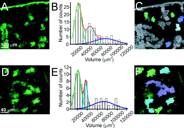
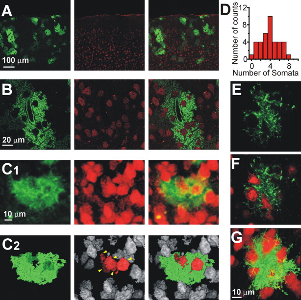
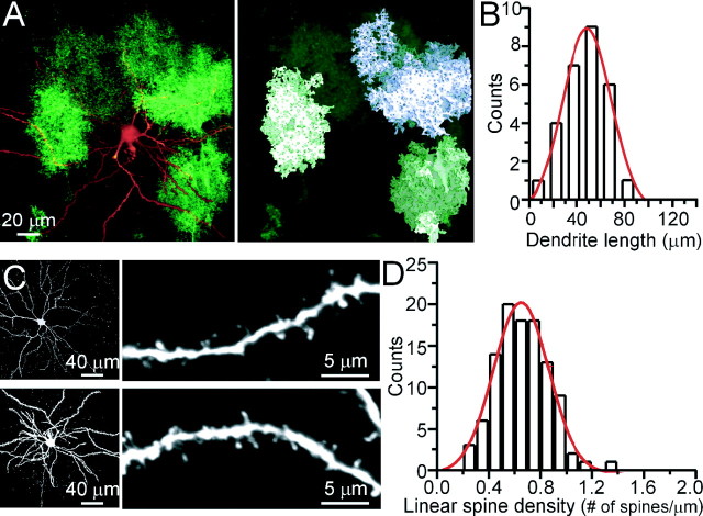
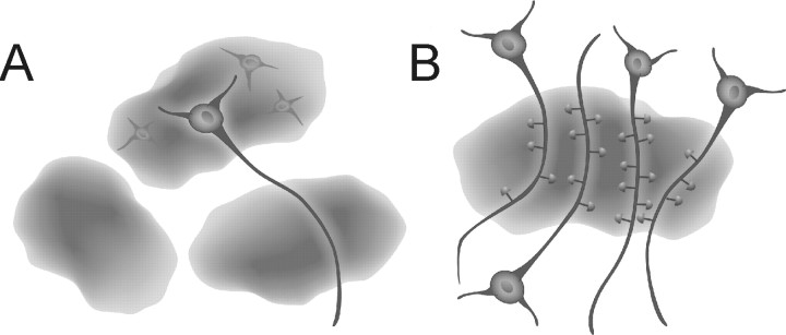
In the mammalian brain, astrocytes modulate neuronal function, in part, by synchronizing neuronal firing and coordinating synaptic networks. Little, however, is known about how this is accomplished from a structural standpoint. To investigate the structural basis of astrocyte-mediated neuronal synchrony and synaptic coordination, the three-dimensional relationships between cortical astrocytes and neurons was investigated. Using a transgenic and viral approach to label astrocytes with enhanced green fluorescent protein, we performed a three-dimensional reconstruction of astrocytes from tissue sections or live animals in vivo. We found that cortical astrocytes occupy nonoverlapping territories similar to those described in the hippocampus. Using immunofluorescence labeling of neuronal somata, a single astrocyte enwraps on average four neuronal somata with an upper limit of eight. Single-neuron dye-fills allowed us to estimate that one astrocyte contacts 300-600 neuronal dendrites. Together with the recent findings showing that glial Ca2+ signaling is restricted to individual astrocytes in vivo, and that Ca2+ signaling leads to gliotransmission, we propose the concept of functional islands of synapses in which groups of synapses confined within the boundaries of an individual astrocyte are modulated by the gliotransmitter environment controlled by that astrocyte. Our description offers a new structurally based conceptual framework to evaluate functional data involving interactions between neurons and astrocytes in the mammalian brain.


Figures

Figure 1.
Figure 1.
Cortical astrocytes occupy nonoverlapping domains. A, Top-view reconstruction of EGFP-expressing cortical astrocytes from dnSNARE coronal brain sections. B, Histogram representing the volume distribution of EGFP labeling of astrocytes. Raw data were fit with three Gaussian curves with peaks at 22,906, 46,188, and 78,684 μm3 (n = 4 dnSNARE mice). C, Based on the analysis performed in B, structures with a volume value within 90% of the first Gaussian function are shown in green, representing single astrocytes. Structures with volumes equivalent to two and three astrocytes are shown in blue and violet, respectively. D, Top-view reconstruction of the EGFP-expressing astrocytes obtained in vivo from a dnSNARE-expressing animal. E, Histogram of astrocyte volume distribution shows three distinct peaks at 21,033, 36,713 and 72,664 (n = 3 dnSNARE animals). F, Using the same criteria and color code as in C, structures corresponding to single, two, and three astrocytes are shown.
Figure 2.
Figure 2.
One astrocyte enwraps several neuronal somata. A and B show individual optical sections to reveal the structure of astrocytes expressing EGFP within the dnSNARE mouse (green; left), the location of neurons immunolabeled with anti-NeuN (red; middle), and an overlay (right). Note that the astrocytes contain several holes, which correspond with the locations of neuronal somata. C1, Top-view reconstruction of a single astrocyte (green) and neurons (red). C2, The same cells shown in C1 are depicted in a top-view reconstruction with a threshold mask. Neurons within the astrocytic territory are red (yellow arrows). D, Distribution of the number of neuronal somata that contact one astrocyte. At least 50% of the surface area of a neuron had to make contact with the astrocyte to be considered as having sufficient interaction for inclusion in this analysis. E, F, A single confocal plane of an AAV-transduced astrocyte (green) contacting neuronal somata (red). G, Three-dimensional reconstruction of the astrocyte in E and F.
Figure 3.
Figure 3.
Single astrocytes enwrap different dendrites of the same neuron. A, Left, Top-view reconstruction showing a biocytin-filled layer 2/3 cortical neuron in a slice from a dnSNARE animal. A volume analysis, performed as in Figure 1, identifies regions of EGFP expression corresponding to one (right, green) or two (violet) astrocytes. B, Bar graph showing the distribution of the dendrite length covered by a single astrocyte. Data are obtained from 24 dendrites of four layer 2/3 cortical neurons from three different animals. C, Left, Two examples of biocytin-filled cortical neurons stained with Alexa-conjugated streptavidin. Right, Higher-magnification images allow the identification of single spines. D, Distribution of the linear density of spines measured in 105 dendrite segments from nine biocytin-filled cortical neurons.
Figure 4.
Figure 4.
Schematic representation of functional synaptic islands. A, Diagram showing three astrocytes. The holes are filled with neurons, one of which is shown to have its processes extending out to other astrocytes pointing to the potential of different neuronal compartments being modulated by different astrocytes. B, Diagram illustrating the concept of functional synaptic islands: a group of dendrites from several neurons are enwrapped by a single astrocyte. Synapses localized within the territory of this astrocyte have the potential to be modulated in a coordinated manner by gliotransmitter(s) released from this glial cell.

References

    1. Angulo MC, Kozlov AS, Charpak S, Audinat E. Glutamate released from glial cells synchronizes neuronal activity in the hippocampus. J Neurosci. 2004;24:6920–6927. - PMC - PubMed
    1. Arcuino G, Lin JH, Takano T, Liu C, Jiang L, Gao Q, Kang J, Nedergaard M. Intercellular calcium signaling mediated by point-source burst release of ATP. Proc Natl Acad Sci USA. 2002 - PMC - PubMed
    1. Ballesteros-Yanez I, Benavides-Piccione R, Elston GN, Yuste R, DeFelipe J. Density and morphology of dendritic spines in mouse neocortex. Neuroscience. 2006;138:403–409. - PubMed
    1. Bezzi P, Carmignoto G, Pasti L, Vesce S, Rossi D, Rizzini BL, Pozzan T, Volterra A. Prostaglandins stimulate calcium-dependent glutamate release in astrocytes. Nature. 1998;391:281–285. - PubMed
    1. Bushong EA, Martone ME, Jones YZ, Ellisman MH. Protoplasmic astrocytes in CA1 stratum radiatum occupy separate anatomical domains. J Neurosci. 2002;22:183–192. - PMC - PubMed

Publication types

MeSH terms

Substances

LinkOut - more resources