. 2007 Jun 13;27(24):6478-88.
doi: 10.1523/JNEUROSCI.0342-07.2007.

Molecular characterization of the ankle-link complex in cochlear hair cells and its role in the hair bundle functioning

Affiliations

Molecular characterization of the ankle-link complex in cochlear hair cells and its role in the hair bundle functioning

Nicolas Michalski et al. J Neurosci. .

Abstract

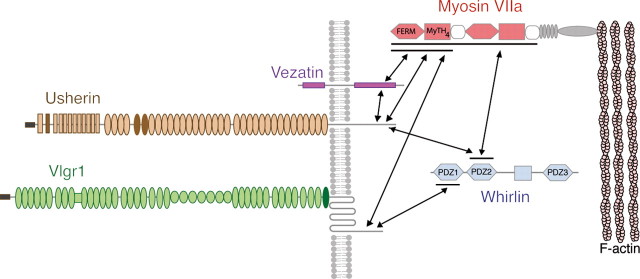
Several lines of evidence indicate that very large G-protein-coupled receptor 1 (Vlgr1) makes up the ankle links that connect the stereocilia of hair cells at their base. Here, we show that the transmembrane protein usherin, the putative transmembrane protein vezatin, and the PDZ (postsynaptic density-95/Discs large/zona occludens-1) domain-containing submembrane protein whirlin are colocalized with Vlgr1 at the stereocilia base in developing cochlear hair cells and are absent in Vlgr1-/- mice that lack the ankle links. Direct in vitro interactions between these four proteins further support their involvement in a molecular complex associated with the ankle links and scaffolded by whirlin. In addition, the delocalization of these proteins in myosin VIIa defective mutant mice as well as the myosin VIIa tail direct interactions with vezatin, whirlin, and, we show, Vlgr1 and usherin, suggest that myosin VIIa conveys proteins of the ankle-link complex to the stereocilia. Adenylyl cyclase 6, which was found at the base of stereocilia, was both overexpressed and mislocated in Vlgr1-/- mice. In postnatal day 7 Vlgr1-/- mice, mechanoelectrical transduction currents evoked by displacements of the hair bundle toward the tallest stereocilia (i.e., in the excitatory direction) were reduced in outer but not inner hair cells. In both cell types, stimulation of the hair bundle in the opposite direction paradoxically resulted in significant transduction currents. The absence of ankle-link-mediated cohesive forces within hair bundles lacking Vlgr1 may account for the electrophysiological results. However, because some long cadherin-23 isoforms could no longer be detected in Vlgr1-/- mice shortly after birth, the loss of some apical links could be involved too. The premature disappearance of these cadherin isoforms in the Vlgr1-/- mutant argues in favor of a signaling function of the ankle links in hair bundle differentiation.


Figures

Figure 1.
Figure 1.
Vlgr1 in normal hair bundle development. Whole-mount preparations of wild-type mouse cochlear middle-coil double labeled for actin (red) and Vlgr1 (green). A, A′, At E17, the Vlgr1 staining of hair cells is located at the base of the emerging hair bundles and extends as a fine peripheral band to the neural edge short microvilli (A, arrowheads). B, B′, At P1, Vlgr1 labeling is restricted to the basal end of the hair bundles both in OHCs (B) and IHCs (B′). C–E, At P4 and P8, in both hair cell types, the Vlgr1 labeling forms a tight band at the base of stereocilia (C–D), which is not detected in Vlgr1−/− mice (E). F, Vlgr1 is no longer detected in P12 Vlgr1+/+ hair bundles. G, The Vlgr1 labeling disappears after BAPTA or subtilisin treatment of P5 cochlear explants. Scale bars, 4 μm.
Figure 2.
Figure 2.
Morphological defects of Vlgr1−/− hair bundles. A–B′, Scanning electron microphotographs of OHC and IHC hair bundles from P6 Vlgr1+/+ and Vlgr1−/− mice. Insets, Enlarged views of stereocilia bases. A, B, Arrowheads point to the ankle links in Vlgr1+/+ hair bundles. A′, B′, Ankle links are absent in Vlgr1−/− hair bundles. C–E′, Scanning electron microphotographs of the surface of the organ of Corti at the apical, middle, and basal coils of cochleas from P6 Vlgr1+/+ and Vlgr1−/− mice. C′, D′, Arrowheads point to neural side supernumerary microvilli in Vlgr1−/− IHCs. Scale bars, 4 μm.
Figure 3.
Figure 3.
Hair bundle distribution of usherin, vezatin, and whirlin in P6 Vlgr1+/+ and Vlgr1−/− cochlear hair cells. A–B′, In Vlgr1+/+ mice, both usherin (A) and vezatin (B) are detected at the base of stereocilia and along the kinocilium. In Vlgr1−/− mice, usherin (A′) and vezatin (B′) labelings are no longer present at the base of the hair bundle but remain in the kinocilium (A′, B′,arrows) both in OHCs and IHCs. C, C′, In Vlgr1+/+ mice, whirlin is detected both at the base and apical end of stereocilia. In Vlgr1−/− mice, only the apical labeling can be seen (C′, arrowheads), both in stereocilia and supernumerary neural edge microvilli. Scale bar, 4 μm.
Figure 4.
Figure 4.
Distribution of Vlgr1, usherin, and vezatin in the apical region of inner hair cells from P6 shaker-1 and whirler mice. In the absence of myosin VIIa (shaker-1 mutant) or whirlin (whirler mutant), the proper hair bundle distributions of Vlgr1, usherin, and vezatin are no longer detected. A–A“, In shaker-1 hair cells, the Vlgr1 labeling forms clumps beneath the apical cell surface (A′), whereas in whirler hair cells, it is detected at the very base of stereocilia, i.e., below the ankle links (A”). B–B“, A faint usherin staining can be seen beneath the apical cell surface in shaker-1 hair cells (B′, arrowheads), whereas usherin cannot be detected in whirler hair cells (B”). C–C“, The vezatin staining forms clumps beneath the apical cell surface in shaker-1 hair cells (C′) and is not detected in whirler hair cells (C”). Scale bar, 4 μm.
Figure 5.
Figure 5.
Myosin VIIa–Vlgr1, myosinVIIa–usherin, and vezatin–usherin in vitro interactions. A, Agarose beads carrying the Myc-tagged vezatin bind to a GST-fusion protein containing the cytoplasmic region of usherin (GST-CytoUsherin) but not to GST alone. B, The myosin VIIa C-terminal MyTH4-FERM fragment (Myosin VIIa Cter) binds to GST-fusion proteins containing the cytoplasmic domain of either Vlgr1 (GST-CytoVlgr1) or usherin (GST-CytoUsherin) but not to GST alone. The positions of the molecular mass markers are indicated on the right.
Figure 6.
Figure 6.
Adenylate cyclase 6 in Vlgr1+/+ and Vlgr1−/− hair bundles. A, Detection of AC transcripts in P6 OHCs by using single-cell RT-PCR. Only AC6 and AC9 transcripts could be detected. B–F, AC6 distribution in the hair bundles of middle-coil cochlear hair cells at different postnatal stages. B, In P1 wild-type mice, AC6 is detected all along OHC stereocilia but not in IHC stereocilia. C, E, In P2 and P7 wild-type OHCs, the AC6 labeling is restricted to the stereocilia base. D, F, In wild-type IHCs, the AC6 labeling can be detected at the stereocilia base from P3. G, H, H′, In P7 Vlgr1−/− OHCs and IHCs, the AC6 labeling is more intense and located all along stereocilia. Scale bars, 4 μm.
Figure 7.
Figure 7.
Mechanoelectrical transduction current recordings in the excitatory direction. A, Examples of MET current recordings in IHCs (red) and OHCs (blue) from Vlgr1+/+ and Vlgr1−/− P7 mice while applying different displacement steps in the excitatory direction (shown on the left) and one 150 nm step in the opposite direction. Cells were voltage clamped at −80 mV. B, Relative Po-displacement curves plotted for Vlgr1+/+ (n = 5) and Vlgr1−/− (n = 8) IHCs (left) and for Vlgr1+/+ (n = 5) and Vlgr1−/− (n = 11) OHCs (right). Relative Po-displacement curves were fit with a normalized second-order Boltzmann function: I = 1/[1 + {exp(a(X0X))}{1 + exp(b(X1X))}], where a = 0.044 nm−1, b = −0.037 nm−1, X0 = 78 nm, X1 = 60.3 nm for Vlgr1+/+ IHCs, and a = 0.024 nm−1, b = −0.017 nm−1, X0 = 123.6 nm, X1 = 86.5 nm for Vlgr1−/− IHCs, and a = 0.049 nm−1, b = −0.036 nm−1, X0 = 80.4 nm, X1 = 46.9 nm for Vlgr1+/+ OHCs, and a = 0.027 nm−1, b = −0.020 nm−1, X0 = 166 nm, X1 = 120.4 nm for Vlgr1−/− OHCs. B′, X0.5 denotes average displacements required to open 50% of the MET channels and average sensitivity is the slope mean value calculated for displacements corresponding to Po values between 0.2 and 0.8 in Vlgr1+/+ and Vlgr1−/− hair cells. Statistically significant differences (Student's t test) are denoted by asterisks.
Figure 8.
Figure 8.
Mechanoelectrical transduction current recordings in the inhibitory direction. A, C, Examples of MET current recordings in IHCs (red) and OHCs (blue) from Vlgr1+/+ and Vlgr1−/− P7 mice while applying different displacement steps in the inhibitory direction. Cells were voltage clamped at −80 mV. B, D, Dihydrostreptomycin (DHS) application suppresses the MET current in P7 Vlgr1−/− IHCs and OHCs.
Figure 9.
Figure 9.
Hair bundle distribution of cadherin 23 in Vlgr1+/+ and Vlgr1−/− inner hair cells. A, B, C, In P1 (A) and P6 (B, C) wild-type hair cells from cochlear middle coil, the N1 (extracellular) and Ela3 (both intracellular and extracellular) epitopes of cadherin 23 are both detected in the apical part of the stereocilia. A′, B′, C′, In Vlgr1−/− hair cells, the N1 epitope is detected at P0 (A′) but not at P6 (B′). In contrast, the distribution of Ela3 is preserved (C′). Scale bar, 4 μm.
Figure 10.
Figure 10.
Schematic representation of proteins involved in the ankle-link complex. In vitro direct interactions between proteins are indicated by arrows.

Similar articles

Cited by

References

    1. Adato A, Kikkawa Y, Reiners J, Alagramam KN, Weil D, Yonekawa H, Wolfrum U, El-Amraoui A, Petit C. Interactions in the network of Usher syndrome type 1 proteins. Hum Mol Genet. 2005a;14:347–356. - PubMed
    1. Adato A, Lefèvre G, Delprat B, Michel V, Michalski N, Chardenoux S, Weil D, El-Amraoui A, Petit C. Usherin, the defective protein in Usher syndrome type IIA, is likely to be a component of interstereocilia ankle links in the inner ear sensory cells. Hum Mol Genet. 2005b;14:3921–3932. - PubMed
    1. Ahmed ZM, Riazuddin S, Bernstein SL, Ahmed Z, Khan S, Griffith AJ, Morell RJ, Friedman TB, Riazuddin S, Wilcox ER. Mutations of the protocadherin gene PCDH15 cause Usher syndrome type 1F. Am J Hum Genet. 2001;69:25–34. - PMC - PubMed
    1. Alagramam KN, Yuan H, Kuehn MH, Murcia CL, Wayne S, Srisailpathy CRS, Lowry RB, Knaus R, Van Laer L, Bernier FP, Schwartz S, Lee C, Morton CC, Mullins RF, Ramesh A, Van Camp G, Hagemen GS, Woychik RP, Smith RJH. Mutations in the novel protocadherin PCDH15 cause Usher syndrome type 1F. Hum Mol Genet. 2001;10:1709–1718. - PubMed
    1. Appert-Collin A, Baisamy L, Diviani D. Regulation of G protein-coupled receptor signaling by A-kinase anchoring proteins. J Recept Signal Transduct Res. 2006;26:631–646. - PubMed

Publication types

MeSH terms