. 2007 Sep;18(9):3366-74.
doi: 10.1091/mbc.e06-10-0895. Epub 2007 Jun 13.

The localization of inner centromeric protein (INCENP) at the cleavage furrow is dependent on Kif12 and involves interactions of the N terminus of INCENP with the actin cytoskeleton

Affiliations

The localization of inner centromeric protein (INCENP) at the cleavage furrow is dependent on Kif12 and involves interactions of the N terminus of INCENP with the actin cytoskeleton

Qian Chen et al. Mol Biol Cell. 2007 Sep.

Abstract

The inner centromeric protein (INCENP) and other chromosomal passenger proteins are known to localize on the cleavage furrow and to play a role in cytokinesis. However, it is not known how INCENP localizes on the furrow or whether this localization is separable from that at the midbody. Here, we show that the association of Dictyostelium INCENP (DdINCENP) with the cortex of the cleavage furrow involves interactions with the actin cytoskeleton and depends on the presence of the kinesin-6-related protein Kif12. We found that Kif12 is found on the central spindle and the cleavage furrow during cytokinesis. Kif12 is not required for the redistribution of DdINCENP from centromeres to the central spindle. However, in the absence of Kif12, DdINCENP fails to localize on the cleavage furrow. Domain analysis indicates that the N terminus of DdINCENP is necessary and sufficient for furrow localization and that it binds directly to the actin cytoskeleton. Our data suggest that INCENP moves from the central spindle to the furrow of a dividing cell by a Kif12-dependent pathway. Once INCENP reaches the equatorial cortex, it associates with the actin cytoskeleton where it then concentrates toward the end of cytokinesis.


Figures

Figure 1.
Figure 1.
Kif12 localizes on the spindle during mitosis and at the cleavage furrow during cytokinesis. Shown are two time-lapse fluorescence images of wild-type or Kif12 null cells expressing GFP–Kif12 during cytokinesis. GFP–Kif12 was found on the mitotic spindle. Moreover, it localized to the cortex of the cleavage furrow (white arrow) during cytokinesis and was clearly seen at the remnant of the intercellular bridge after the daughter cells separated (see Supplemental Movie 1). Times are indicated in minutes:seconds. Bar, 5 μm.
Figure 2.
Figure 2.
Kif12 is essential for the redistribution of DdINCENP from the central spindle to the cortex of the cleavage furrow. Fluorescence microscopy images of cells expressing GFP–DdINCENP during cytokinesis. (A and B) GFP–DdINCENP is normally found at the spindle poles and central spindle as seen in these dividing wild-type cells. During cytokinesis, GFP–DdINCENP then localizes on the cortex of the cleavage furrow (arrows) (see Supplemental Movie 2). (C and D) GFP–DdINCENP is still localized on the central spindle and spindle poles in Kif12 null cells. However, GFP– DdINCENP failed to localize at the cleavage furrow in the absence of Kif12 (arrows). (E) A time-lapse series of a DdKif12 null cell expressing GFP–DdINCENP shows that GFP–DdINCENP was never found at the cleavage furrow during cytokinesis. Arrows point to the cleavage furrow. Noticeably, GFP–DdINCENP was also absent from the cytoplasmic bridge at the end of cytokinesis, whereas it highly concentrated on the bridge in wild-type cells (see Supplemental Movie 3). Times are indicated in minutes:seconds. Bar, 5 μm.
Figure 3.
Figure 3.
The localization of Kif12 during mitosis and cytokinesis does not depend on DdINCENP. Time-lapse fluorescence micrographs of DdINCENP null cells expressing GFP-DdKif12. (A) GFP-Kif12 still localized on the mitotic spindle (arrowheads) in an DdINCENP null cell. The cell was entering cytokinesis when the central spindle dismantled and GFP–Kif12 began to accumulate at the cortex of the cleavage furrow (arrows). (B) In DdINCENP null cells, GFP–Kif12 was still found at the cortex of the cleavage furrow (arrows) at the early stage of cytokinesis and continued to build up at the furrow area. At the end of the cytokinesis, GFP–Kif12 was highly enriched at the cytoplasmic bridge (see Supplemental Movie 4). Note that the central spindle was out of focus in most frames in this movie. This focal plane helps highlight the association of GFP–Kif12 with the ingressing cleavage furrow. Times are indicated in minutes:seconds. Bar, 5 μm.
Figure 4.
Figure 4.
Domain analysis of DdINCENP. DdINCENP contains an IN-box domain near its C terminus, two coiled coil domains, and a single nuclear localization signal (*) at position 728–755. Shown are the four truncated mutants of DdINCENP used in this study. The IN-box is the only region with recognizable similarities among different INCENPs. The IN-box domain of DdINCENP is 44% identical to that of fission yeast INCENP and 21% identical to that of chicken INCENP (Chen et al., 2006).
Figure 5.
Figure 5.
The N terminus of DdINCENP is essential for its localization at the cleavage furrow during cytokinesis. The four different truncated mutants of DdINCENP were tagged with GFP and expressed in DdINCENP null cells. Time-lapse fluorescence micrographs of these cells during cytokinesis are shown. The mutant protein that lacks the N terminus, DdINCENP488-1320, was absent from the cortex of the cleavage furrow (arrows), and it was homogenously distributed in the cytoplasm during cytokinesis. In contrast, the three proteins that contain the N terminus, DdINCENP1-1013, DdINCENP1-500, and DdINCENP1-273, were all found at the cortex of the cleavage furrow during cytokinesis (arrows) (see Supplemental Movies 5–8). The focal planes shown here do not display the association of these proteins with the spindle poles and central spindles, but they are clearly shown in Figure 6. Times are indicated in minutes:seconds. Bar, 5 μm.
Figure 6.
Figure 6.
Localization of DdINCENP at the central spindle does not depend on either the IN-box or its N-terminal domain. The GFP-tagged DdINCENP mutants were expressed in DdINCENP null cells. Fluorescence microscopy images of these cells during anaphase are shown. (A–E) The GFP fluorescence signal is shown. (A′–E′) Merged fluorescence micrographs to show DNA (blue) and GFP fluorescence (green). All four DdINCENP mutants localized to the spindle pole bodies and the central spindles (arrows) during anaphase. GFP–DdINCENP1-1013 had the brightest signal on the central spindle compared with the proteins (C, C′, D, and D′). Additionally, GFP–DdINCENP1-1013 localized to the nuclear envelope (arrow heads) during telophase (D). Bar, 5 μm.
Figure 7.
Figure 7.
DdINCENP1-500 associates with the actin cytoskeleton during interphase. (A) Time-lapse fluorescence micrographs of wild-type cells (WT) expressing GFP–DdINCENP1-500 are shown. The protein was enriched at the bottom portion of pinocytic cups (arrows) during endocytosis (see Supplemental Movie 9). (B, B′, and B″) Wild-type cells expressing GFP–DdINCENP1-500 were fixed and immunostained with Texas Red-labeled phalloidin to visualize F-actin (B). The chimera protein was colocalized with F-actin on the pinocytic cups (arrowheads). Times are indicated in minutes:seconds. Bar, 5 μm.
Figure 8.
Figure 8.
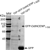
TAP-tagged GFP–DdINCENP1-500 associates with components of the actin cytoskeleton. TAP–GFP–DdINCENP1-500 and TAP–GFP were expressed separately in wild-type cells and purified by binding to IgG beads followed by cleavage elution with TEV protease. The purified protein samples were loaded on the SDS-PAGE gel, which was stained with Coomassie blue. Comparison of the protein profile identified four proteins that specifically copurified with TAP—GFP–DdINCENP1-500. These four proteins were identified by mass spectrometry as actin (a), ABP-50 (b), Hsp-70 (c), and myosin II heavy chain (d).

Similar articles

Cited by

References

    1. Abaza A., Soleilhac J. M., Westendorf J., Piel M., Crevel I., Roux A., Pirollet F. M phase phosphoprotein 1 is a human plus-end-directed kinesin-related protein required for cytokinesis. J. Biol. Chem. 2003;278:27844–27852. - PMC - PubMed
    1. Adams R. R., Carmena M., Earnshaw W. C. Chromosomal passengers and the (aurora) ABCs of mitosis. Trends Cell Biol. 2001a;11:49–54. - PubMed
    1. Adams R. R., Maiato H., Earnshaw W. C., Carmena M. Essential roles of Drosophila inner centromere protein (INCENP) and Aurora B in histone H3 phosphorylation, metaphase chromosome alignment, kinetochore disjunction, and chromosome segregation. J. Cell Biol. 2001b;153:865–880. - PMC - PubMed
    1. Adams R. R., Tavares A. A., Salzberg A., Bellen H. J., Glover D. M. pavarotti encodes a kinesin-like protein required to organize the central spindle and contractile ring for cytokinesis. Genes Dev. 1998;12:1483–1494. - PMC - PubMed
    1. Adams R. R., Wheatleya S. P., Gouldsworthy A. M., Kandels-Lewis S. E., Carmena M., Smythe C., Gerloff D. L., Earnshaw W. C. INCENP binds the Aurora-related kinase AIRK2 and is required to target it to chromosomes, the central spindle and cleavage furrow. Curr. Biol. 2000;10:1075–1078. - PubMed

Publication types

LinkOut - more resources