. 2007 Jun;17(6):947-53.
doi: 10.1101/gr.6073107.

Construction of a genome-scale structural map at single-nucleotide resolution

Affiliations

Construction of a genome-scale structural map at single-nucleotide resolution

Jason A Greenbaum et al. Genome Res. 2007 Jun.

Abstract

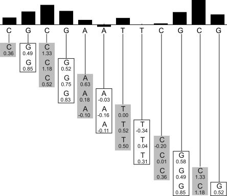
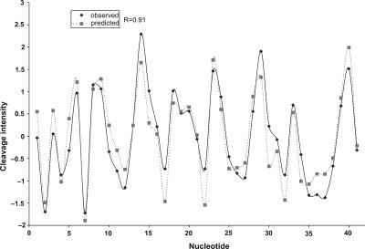
Few methods are available for mapping the local structure of DNA throughout a genome. The hydroxyl radical cleavage pattern is a measure of the local variation in solvent-accessible surface area of duplex DNA, and thus provides information on the local shape and structure of DNA. We report the construction of a relational database, ORChID (OH Radical Cleavage Intensity Database), that contains extensive hydroxyl radical cleavage data produced from two DNA libraries. We have used the ORChID database to develop a set of algorithms that are capable of predicting the hydroxyl radical cleavage pattern of a DNA sequence of essentially any length, to high accuracy. We have used the prediction algorithm to produce a structural map of the 30 Mb of the ENCODE regions of the human genome.


Figures

Figure 1.
Figure 1.
(A) Design of the DNA molecules used to construct the R40 library. The test sequence (an insert of 40 random nucleotides) is located near the center of the DNA strand, flanked by common sequences on both sides. The pentamer library was constructed in a similar manner, with individual pentamer sequences (see Supplemental Fig. S1) serving as the test sequence. (B) Typical electropherogram of a sample from the R40 library. The R40 test sequence is boxed, and the common flanking sequences are shaded. Each peak in the pattern represents cleavage by the hydroxyl radical at one nucleotide of the DNA molecule. The area of a peak is proportional to the extent of cleavage at that nucleotide (Shadle et al. 1997). Note that the cleavage patterns of the common palindromic flanking sequences at the 5′ (left) and 3′ (right) ends are similar for a particular member of the library. This also holds true for different library members (data not shown).
Figure 2.
Figure 2.
Reproducibility of the cleavage pattern of the common flanking sequence (see Fig. 1). The mean cleavage intensities, taken from 112 instances of the common flanking sequence cleavage pattern, are plotted as bars, with standard deviations shown as error bars.
Figure 3.
Figure 3.
Cleavage/sequence correlation at various levels of sequence identity. Heat maps were created with Matrix2png (Pavlidis and Noble 2003), using the data from Supplemental Tables S5, S6, and S7 as input. (A) 10-mers; (B) 20-mers; (C) 30-mers. The intensity of each rectangle indicates the number of pairs of sequences in that cell, ranging from black (lowest) to white (highest).
Figure 4.
Figure 4.
High similarity in cleavage patterns for two sequences with low sequence identity. Plotted are the hydroxyl radical cleavage patterns of two 10-mer sequences that share no common nucleotides (sequence identity = 0%). Note the significant correlation (R = 0.94) of the two patterns. (See Supplemental Table S8 for sequences.)
Figure 5.
Figure 5.
Sliding trimer window algorithm. The sequence to be predicted is shown below the bar graph. It is divided into overlapping trinucleotides. The hydroxyl radical cleavage intensity data for each trimer are retrieved from the ORChID database and are listed below each nucleotide. The values in each column are averaged to produce a predicted hydroxyl radical cleavage intensity, which is represented as a bar at the top. Note that the two terminal nucleotides at each end rely on data from only one or two trinucleotides, rather than three.
Figure 6.
Figure 6.
Predicted hydroxyl radical cleavage pattern. The hydroxyl radical cleavage pattern of sample ID 25201 from the ORChID database was predicted using the Sliding Trimer Window algorithm (see Fig. 5). The predicted pattern (broken line) is compared to the experimental pattern (solid line). The experimental and predicted patterns have a correlation coefficient of 0.91.

References

    1. Angermayr M., Oechsner U., Gregor K., Schroth G.P., Bandlow W., Oechsner U., Gregor K., Schroth G.P., Bandlow W., Gregor K., Schroth G.P., Bandlow W., Schroth G.P., Bandlow W., Bandlow W. Transcription initiation in vivo without classical transactivators: DNA kinks flanking the core promoter of the housekeeping yeast adenylate kinase gene, AKY2, position nucleosomes and constitutively activate transcription. Nucleic Acids Res. 2002;30:4199–4207. - PMC - PubMed
    1. Balasubramanian B., Pogozelski W.K., Tullius T.D., Pogozelski W.K., Tullius T.D., Tullius T.D. DNA strand breaking by the hydroxyl radical is governed by the accessible surface areas of the hydrogen atoms of the DNA backbone. Proc. Natl. Acad. Sci. 1998;95:9738–9743. - PMC - PubMed
    1. Barbic A., Zimmer D.P., Crothers D.M., Zimmer D.P., Crothers D.M., Crothers D.M. Structural origins of adenine-tract bending. Proc. Natl. Acad. Sci. 2003;100:2369–2373. - PMC - PubMed
    1. Berman H.M. Crystal studies of B-DNA: The answers and the questions. Biopolymers. 1997;44:23–44. - PubMed
    1. Berman H.M., Olson W.K., Beveridge D.L., Westbrook J., Gelbin A., Demeny T., Hsieh S.H., Srinivasan A.R., Schneider B., Olson W.K., Beveridge D.L., Westbrook J., Gelbin A., Demeny T., Hsieh S.H., Srinivasan A.R., Schneider B., Beveridge D.L., Westbrook J., Gelbin A., Demeny T., Hsieh S.H., Srinivasan A.R., Schneider B., Westbrook J., Gelbin A., Demeny T., Hsieh S.H., Srinivasan A.R., Schneider B., Gelbin A., Demeny T., Hsieh S.H., Srinivasan A.R., Schneider B., Demeny T., Hsieh S.H., Srinivasan A.R., Schneider B., Hsieh S.H., Srinivasan A.R., Schneider B., Srinivasan A.R., Schneider B., Schneider B. The nucleic acid database. A comprehensive relational database of three-dimensional structures of nucleic acids. Biophys. J. 1992;63:751–759. - PMC - PubMed

Publication types

LinkOut - more resources