. 2007 May-Jun;27(3):161-5.
doi: 10.5144/0256-4947.2007.161.

Biliary ascariasis: the value of ultrasound in the diagnosis and management

Affiliations

Biliary ascariasis: the value of ultrasound in the diagnosis and management

Mohammad Al Absi et al. Ann Saudi Med. 2007 May-Jun.

Abstract

Background: Conventional methods of radiographic examination are often unsatisfactory for identifying worms in the biliary tract. Ultrasonography is a non-invasive, quick and safe procedure known to have diagnostic accuracy. We studied the ultrasonographic appearances of biliary ascariasis and the role of ultrasonography in diagnosis and management.

Methods: In a prospective 5-year study, a sonographic diagnosis of biliary ascariasis was made on 46 Yemeni patients. The diagnosis was based mainly on sonographic appearances supported by clinical and laboratory results and proved by outcome of either surgical or medical management or spontaneous exit of worms. Follow-up ultrasound was performed for all patients to confirm the diagnosis and to monitor management.

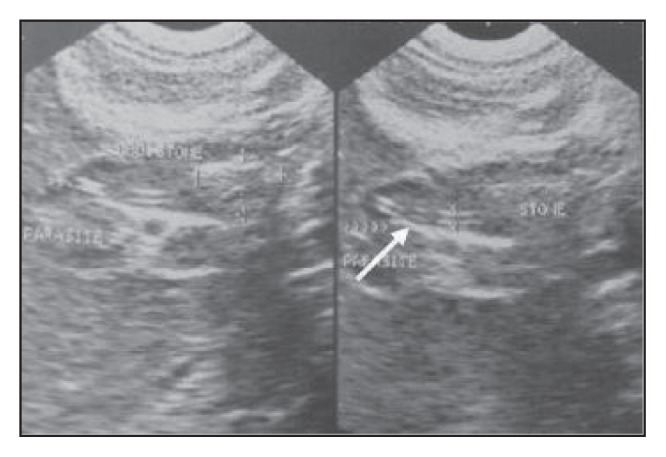
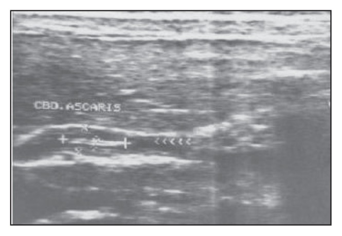
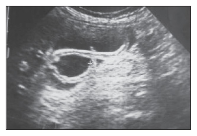
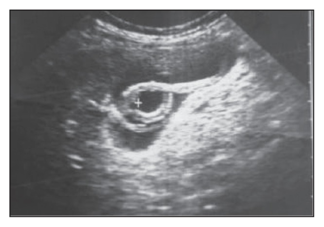
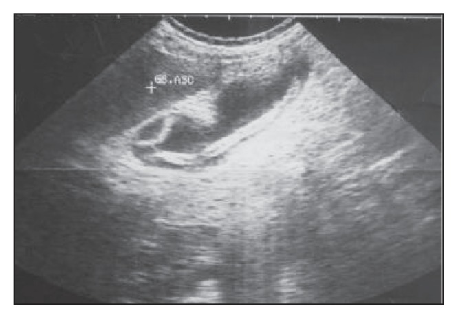
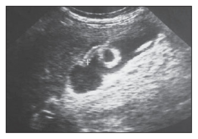
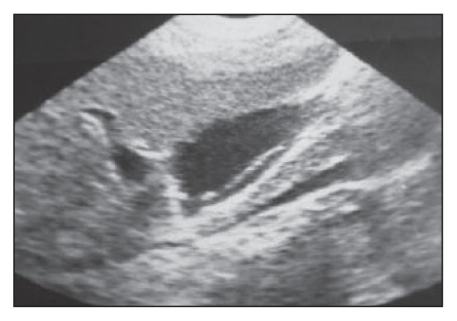
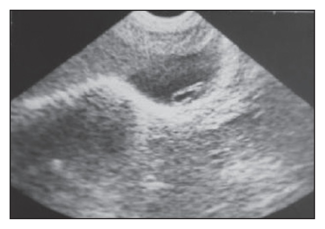
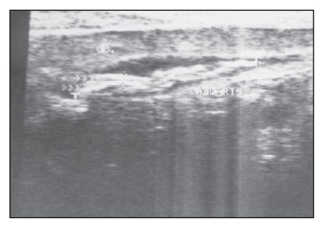
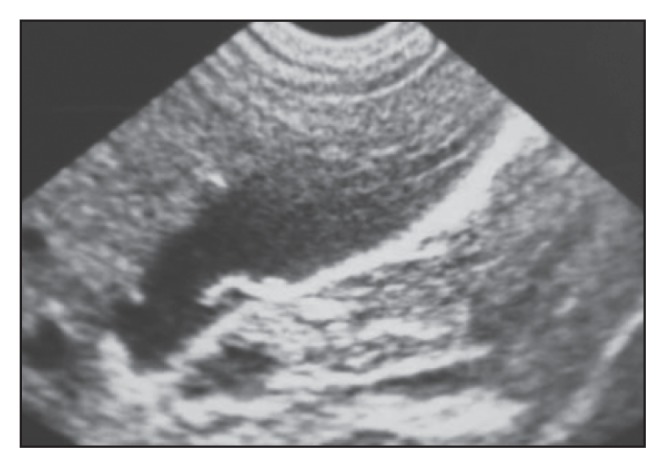
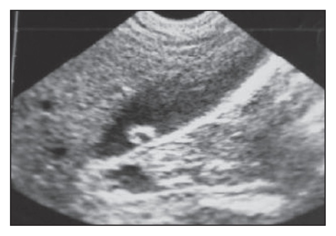
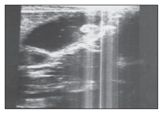
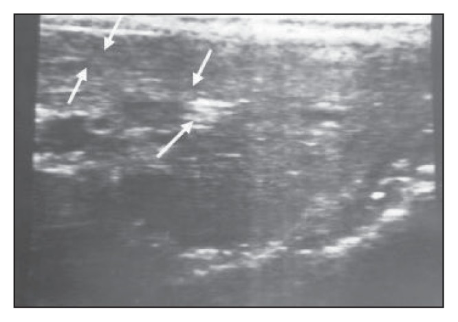
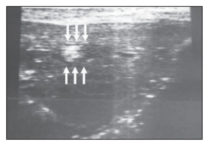
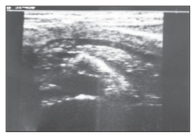
Results: Parasites were present in the dilated main bile duct in 23 patients, in the gallbladder in 12 patients, in the intrahepatic ducts in 6 patients, in the main pancreatic duct in 4 patients and as an intrahepatic abscess in one patient. The characteristic appearance of Ascaris lumbricoides was as single or multiple echogenic non- shadowing linear or curved strips with or without echoic tubular central lines that represent the digestive tracts of the worm. A spaghetti-like appearance was seen in 9 patients and amorphous fragments were seen in 2 patients. Sixteen patients underwent surgery, 20 patients were treated medically (including spontaneous exit of the worm in 7 patients without treatment) and in 10 patients worms were extracted by endoscopic retrograde cholangiopancreatography.

Conclusions: Follow-up ultrasound was found to be effective in confirming the diagnosis and monitoring management.


Figures

Figure 1
Figure 1
An oblique ultrasound scan of a 25-year-old female patient demonstrating fusiform expansion of the common bile duct (at surgery proved to be a choledochal cyst), which contains a rather large shadowing stone with an associated echogenic non-shadowing tubular structure (with anechoic linear center) proximal to the stone (arrow) and extending to the common hepatic duct.
Figure 2
Figure 2
Another female patient aged 16 years with fusiform expansion of the common bile duct containing a non-shadowing, echogenic tubular structure with an anechoic linear center that showed active movement during examination.
Figure 3a
Figure 3a
Longitudinal ultrasound section of a relatively distended gallbladder containing a long echogenic, non-shadowing intraluminal tubular structure with a central anechoic linear area (the “inner tube” sign), representing the digestive tract of the worm.
Figure 3b
Figure 3b
Follow-up of Figure 3a shows change in configuration of the previously seen worm with the appearance of an additional smaller one posterior to it.
Figure 4a
Figure 4a
Ultrasound scan of a gallbladder of another patient showing two intralumenal worms.
Figure 4b
Figure 4b
Change in the configuration and positioning of the worm seen in Figure 4a on the control ultrasound, confirming the diagnosis.
Figure 5a
Figure 5a
A longitudinal section of gallbladder with the worm represented as an echogenic, non-shadowing tube-like structure (strip sign).
Figure 5b
Figure 5b
Change in configuration of the worm in Figure 5a. with the worm folded on itself on the control scan, confirming the diagnosis.
Figure 6
Figure 6
A longitudinal scan of a relatively contracted gallbladder showing several non-shadowing, overlapped, tube-like worms (“spaghetti” sign).
Figure 7a
Figure 7a
In this patient, the gallbladder shows short non-shadowing hyperchoic curved strips that become ring-like in Figure 7b.
Figure 7b
Figure 7b
The curved strips in Figure 7a have become ring-like.
Figure 8
Figure 8
A longitudinal section of the gallbladder of a 15-year-old female patient shows a comma-shaped worm that exhibits active movement during ultrasound examination.
Figure 9a
Figure 9a
A transverse scan through the left hepatic lobe shows a dilated intrahepatic duct that is packed with worms, producing an amorphous appearance and some worms in the form of a bolus.
Figure 9b
Figure 9b
Another transverse scan through the left hepatic lobe showing a dilated intrahepatic duct packed with worms in the form of a bolus.
Figure 10
Figure 10
An ultrasound scan through the pancreas showing a worm bolus in the proximal segment of the dilated pancreatic duct.

Similar articles

Cited by

References

    1. Gabaldon A, Mofidi C, Morishita K, et al. Control of Ascariasis. Report of WHO expert Committee. World Health Organ. Tech, Report Ser. 1967;379:6–7.
    1. Crompton DWT. The Prevalence of Ascaris. Parasitol Today. 1988;4:162–8. - PubMed
    1. . Ascariasis (editorial) Lancet. 1989;1:997–998. - PubMed
    1. Khuroo MS, Zarger SA, Yattoo GN, et al. Ascariasis Indicated Acute Pancreatitis. Br J Surg. 1992;79:1335–8. - PubMed
    1. Gomez NA, Leon CJ, Ortiz O. Ultrasound in the Diagnosis of Round Worms in Gallbladder and Common Bile Duct. Surg Endosc. 1993 Jul-Aug;7(4):339–42. - PubMed

LinkOut - more resources