. 2007 Jun 14;447(7146):864-8.
doi: 10.1038/nature05859.

RAS-RAF-MEK-dependent oxidative cell death involving voltage-dependent anion channels

Affiliations

RAS-RAF-MEK-dependent oxidative cell death involving voltage-dependent anion channels

Nicholas Yagoda et al. Nature. .

Abstract

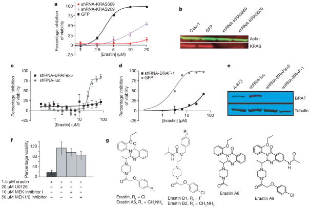
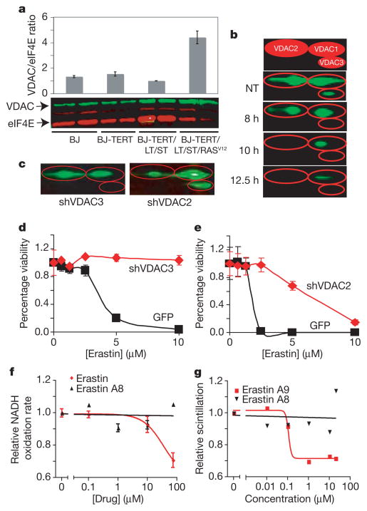
Therapeutics that discriminate between the genetic makeup of normal cells and tumour cells are valuable for treating and understanding cancer. Small molecules with oncogene-selective lethality may reveal novel functions of oncoproteins and enable the creation of more selective drugs. Here we describe the mechanism of action of the selective anti-tumour agent erastin, involving the RAS-RAF-MEK signalling pathway functioning in cell proliferation, differentiation and survival. Erastin exhibits greater lethality in human tumour cells harbouring mutations in the oncogenes HRAS, KRAS or BRAF. Using affinity purification and mass spectrometry, we discovered that erastin acts through mitochondrial voltage-dependent anion channels (VDACs)--a novel target for anti-cancer drugs. We show that erastin treatment of cells harbouring oncogenic RAS causes the appearance of oxidative species and subsequent death through an oxidative, non-apoptotic mechanism. RNA-interference-mediated knockdown of VDAC2 or VDAC3 caused resistance to erastin, implicating these two VDAC isoforms in the mechanism of action of erastin. Moreover, using purified mitochondria expressing a single VDAC isoform, we found that erastin alters the permeability of the outer mitochondrial membrane. Finally, using a radiolabelled analogue and a filter-binding assay, we show that erastin binds directly to VDAC2. These results demonstrate that ligands to VDAC proteins can induce non-apoptotic cell death selectively in some tumour cells harbouring activating mutations in the RAS-RAF-MEK pathway.


Figures

Figure 1
Figure 1. Erastin activates a rapid, oxidative, non-apoptotic cell death process
a, HRASV12-expressing cell lines are sensitive to erastin, whereas isogenic lines lacking HRASV12 are resistant, as determined by Trypan blue exclusion; the graph is a representative outcome of multiple independent experiments. b, Transmission electron microscopy images (×20,000) of BJ-TERT/LT/ST/RASV12 mitochondria after cells were treated with nothing, erastin (37 μM for 10 h) or staurosporine (STS, 1 μM for 5 h). c, Phase-contrast photograph of BJ-TERT/LT/ST/RASV12 cells 24 h after 9 μM erastin treatment indicates that nuclei are intact after cell death. d, Anti-oxidants suppress erastin-induced death in BJ-TERT/LT/ST/RASV12 cells. BHT, butylated hydroxytoluene; DMSO, dimethylsulphoxide. e, Level of intracellular oxidative species in BJ-TERT/LT/ST/RASV12 (black line) or BJ-TERT (grey line) cells treated with 4.6 μM erastin. y axis, percentage of cells exhibiting dichlorofluorescein (DCF) fluorescence within region of interest, M1, as measured by flow cytometry; error bars, one standard deviation, n = 2. f, PARP1 cleavage is not seen during erastin-induced cell death in A-673, HT-1080 and HeLa cells. NT, no treatment. g, STS, but not erastin, induces cytochrome c (cyt c) release from BJ-TERT/LT/ST/RASV12 mitochondria; mitochondrial fraction, mito; cytosolic fraction, cyto. h, Pro-caspase-3 is not cleaved in response to erastin.
Figure 2
Figure 2. Erastin lethality is dependent on the RAS–RAF–MEK pathway
a, Calu-1 cells infected with lentivirus containing shRNAs targeting KRAS were resistant to erastin-induced lethality as quantified by way of comparison with green fluorescent protein (GFP) control in Trypan blue exclusion assay. Sequences of shRNAs are indicated by starting nucleotide in the KRAS mRNA coding sequence. b, KRAS knockdown was confirmed by western blot analysis. c, d, A-673 cells were infected with lentivirus expressing indicated shRNAs or a control GFP plasmid. Cells were treated with erastin for 24 h; percentage inhibition of viability (y axis) was measured using c, Alamar blue and d, Trypan blue. Luc, luciferase; BRAFex5, exon 5 of BRAF transcript. e, BRAF knockdown was confirmed by western blot analysis. f, 48 h treatment with MEK inhibitors (U0126, Sigma; MEK inhibitor 1, Calbiochem; MEK1/2 inhibitor, Calbiochem) prevents erastin-induced lethality in BJ-TERT/LT/ST/RASV12 cells; percentage viability (y axis) was determined using Trypan blue. g, Structures of erastin and related analogues. All error bars in Fig. 2 represent one standard deviation; n =2 or 3.
Figure 3
Figure 3. Erastin compounds act through VDACs
a, VDAC/eukaryotic initiation factor 4E (eIF4E) protein ratio in engineered BJ-derived cells as quantified using western blot. b, BJ-TERT/LT/ST/RASV12 cells were treated with erastin and harvested at indicated time points for quantitative two-dimensional western blot. The top panel is an illustration of VDAC isoforms separated by two-dimensional gel electrophoresis. c, Isoform-specific knockdown of VDAC in HT-1080 cells infected with virus expressing either VDAC3- or VDAC2-targeted shRNA plasmid (shVDAC3 or shVDAC2) was confirmed using two-dimensional protein gels. d, e, These cells were then treated with erastin dilutions, and viability relative to no treatment (y axis) was determined using Trypan blue exclusion and compared to an identical process using a GFP control plasmid. f, Rate of NADH oxidation, normalized to no treatment, in mitochondria purified from VDAC-knockout yeast expressing murine VDAC2 was determined in the presence of erastin (red) or an inactive analogue, erastin A8 (black). g, Direct binding to VDAC2 was assayed using tritium-labelled erastin A9 in competition with unlabelled erastin A9 (red) or erastin A8 (black). All error bars in Fig. 3 represent one standard deviation, n =2 or 3.

References

    1. Kaelin WG. The concept of synthetic lethality in the context of anticancer therapy. Nature Rev Cancer. 2005;5:689–698. - PubMed
    1. Hahn WC, et al. Creation of human tumour cells with defined genetic elements. Nature. 1999;400:464–468. - PubMed
    1. Dolma S, Lessnick SL, Hahn WC, Stockwell BR. Identification of genotype-selective antitumor agents using synthetic lethal chemical screening in engineered human tumor cells. Cancer Cell. 2003;3:285–296. - PubMed
    1. Root DE, Flaherty SP, Kelley BP, Stockwell BR. Biological mechanism profiling using an annotated compound library. Chem Biol. 2003;10:881–892. - PubMed
    1. Song Z, Steller H. Death by design: mechanism and control of apoptosis. Trends Cell Biol. 1999;9:M49–M52. - PubMed

MeSH terms