. 2007 Jun 14;447(7146):869-74.
doi: 10.1038/nature05877.

Resolvin E1 and protectin D1 activate inflammation-resolution programmes

Affiliations

Resolvin E1 and protectin D1 activate inflammation-resolution programmes

Jan M Schwab et al. Nature. .

Abstract

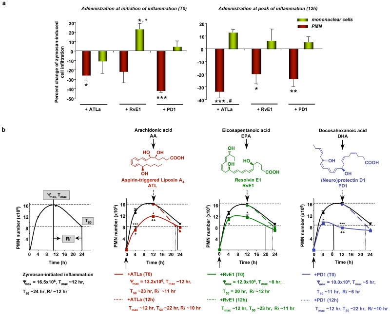
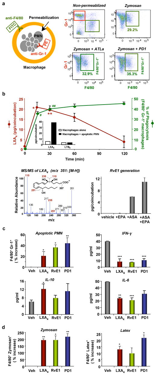
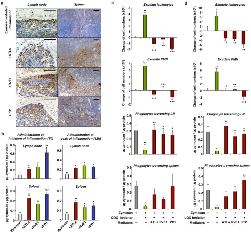
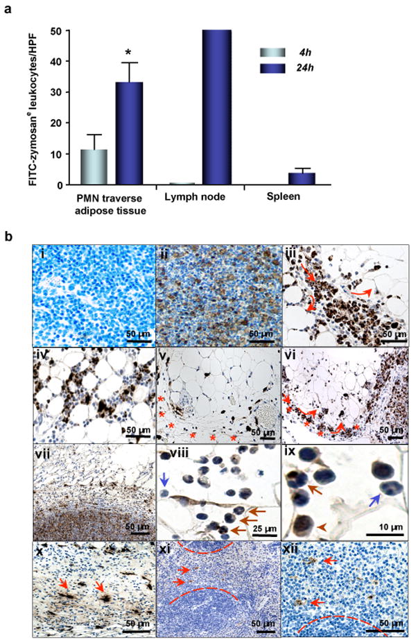
Resolution of acute inflammation is an active process essential for appropriate host responses, tissue protection and the return to homeostasis. During resolution, specific omega-3 polyunsaturated fatty-acid-derived mediators are generated within resolving exudates, including resolvin E1 (RvE1) and protectin D1 (PD1). It is thus important to pinpoint specific actions of RvE1 and PD1 in regulating tissue resolution. Here we report that RvE1 and PD1 in nanogram quantities promote phagocyte removal during acute inflammation by regulating leukocyte infiltration, increasing macrophage ingestion of apoptotic polymorphonuclear neutrophils in vivo and in vitro, and enhancing the appearance of phagocytes carrying engulfed zymosan in lymph nodes and spleen. In this tissue terrain, inhibition of either cyclooxygenase or lipoxygenases--pivotal enzymes in the temporal generation of both pro-inflammatory and pro-resolving mediators--caused a 'resolution deficit' that was rescued by RvE1, PD1 or aspirin-triggered lipoxin A4 analogue. Also, new resolution routes were identified that involve phagocytes traversing perinodal adipose tissues and non-apoptotic polymorphonuclear neutrophils carrying engulfed zymosan to lymph nodes. Together, these results identify new active components for postexudate resolution traffic, and demonstrate that RvE1 and PD1 are potent agonists for resolution of inflamed tissues.


Figures

Figure 1
Figure 1. Pro-resolving lipid mediators RvE1, PD1 and ATLa direct local phagocyte tissue flux
(A) Peritonitis. ATLa, RvE1, or PD1 (300 ng, ip) was given either with zymosan (T0, left) or after (12h, right), and peritoneal exudates collected (24h). Results are the mean ± s.e.m (n=4-8). *p<0.05, **p<0.01, ***p<0.001, compared to zymosan alone; +p<0.05, ATLa vs. RvE1; #p<0.05, T0 vs. 12h (B) Resolution Indices were defined in ref. 12 ( Supplemental Fig. 1), including ψmax (maximal PMN), Tmax (time point when PMNs reach ψmax), T50 (time point corresponding to ∼50% PMN reduction) and Ri (resolution interval, the interval between Tmax and T50). These indices were calculated when compounds were given at the initiation (T0, solid lines and arrows) or peak (12h, dashed lines and arrows) of inflammation. Results are the mean ± s.e.m. (n=4-8). *p<0.05, ***p<0.001, compared to zymosan alone; +p<0.05, ATLa vs. RvE1; ++p<0.01, ATLa vs. PD1.
Figure 2
Figure 2. RvE1 and PD1 increase macrophage phagocytic activity in vivo and in vitro
(A) In vivo phagocytosis with representative dot plots of FACS analysis. (B) (Top) Time course of eicosanoid generation and phagocytosis activity in vitro. Results are the mean ± s.e.m. (n=3). LXA4 amounts are expressed as pg/coincubation (0.4x10⁶ PMN+0.2x10⁶ macrophages in 0.2 ml). *p=0.01, **p=0.02 (vs. time 0 or 120 min). Phagocytosis activities are expressed as [F4/80+Gr-1+/F4/80+]x100%. #p=0.03, ##p=0.01 (vs. time 120 min). (Inset) LXA4 and LTB4 generation at 60 min. (Bottom left) MS/MS spectrum of LXA4. (Bottom right) RvE1 generation. Macrophages were incubated with ASA (500 μM, 30 min) followed by EPA (20 μM, 45 min) prior to incubation with apoptotic PMN. RvE1 was determined by LC/MS/MS and expressed as pg/coincubation (2x10⁶ PMN+1x10⁶ macrophages in 1 ml). (C) Phagocytosis of apoptotic PMN in vitro. Results are the mean ± s.e.m. (n=3-4) and expressed as percent increase of F4/80+Gr-1+ macrophages compared to vehicle alone. Cytokine/chemokine levels are the mean ± s.e.m. (n=3). *p<0.05, **p<0.01, ***p<0.001, compared to vehicle alone. (D) Phagocytosis of zymosan and latex particles in vitro. Results are the mean ± s.e.m. (n=3) and expressed as percent increase of F4/80+zymosan+ or F4/80+latex+ macrophages compared to vehicle alone. Results are the mean ± s.e.m. (n=3). *p<0.05, **p<0.01 compared to vehicle alone.
Figure 3
Figure 3. RvE1 and PD1 enhance leukocytes carrying phagocytozed zymosan in lymph nodes and spleen
(A) Leukocytes with engulfed-zymosan particles (zymosane) in the cortex of the LN and marginal zone of the spleen. The brown particles are positive staining of zymosan. Scale bars: 50 μm. (B) Quantification of zymosan. ATLa, RvE1 or PD1 was given at initiation (T0) or peak (12 h) of inflammation. Results are mean ± s.e.m. (n=4-8). *p=0.05, **p<0.01, ***p<0.001. (C, D) COX or LOX inhibitor was given 30 min before zymosan challenge with or without mediators. Results are mean ± s.e.m. (n=3-4). *p=0.05, **p<0.01, ***p<0.001, compared with zymosan alone; +p=0.05, ++p<0.01, +++p<0.001, compared with (C) zymosan + COX-inhibitor or (D) zymosan + LOX-inhibitor.
Figure 4
Figure 4. Active removal of leukocytes from the inflammatory exudate
(A) Quantitation of FITC-zymosane leukocytes. Results are mean ± s.e.m. (n=3). *p=0.05 (4h vs. 24h). (B) Tracking of FITC-zymosane leukocytes 24h after FITC-zymosan injection in the (i) absence or (ii) presence of the primary anti-FITC Ab. The brown particles are positive staining of FITC-zymosan. (iii) Lipopassage (arrows). (iv) Zymosane leukocytes in perinodal adipose tissue. (v) In perinodal adipose tissue, lymph vessels (stars) conduct zymosane leukocytes in proximity to blood vessels. (vi) Zymosane leukocytes in the afferent lymph vessel (stars) and subcapsular sinus. (vii) In the lymph nodes, zymosane leukocytes were detected markedly in cortex areas. (viii, ix) Zymosane leukocytes consisted of monocytes/macrophages (arrowheads), and non-apoptotic PMNs with engulfed-zymosan (brown arrows) or without (blue arrows). (x) Dendritic-like (fine cell protrusions), zymosane leukocytes in the afferent lymph vessels (arrows). (xi, xii) In the spleen, some zymosane leukocytes also displaced morphological characteristics of DCs (arrows).

References

    1. Serhan CN, Savill J. Resolution of inflammation: the beginning programs the end. Nat Immunol. 2005;6:1191–1197. - PubMed
    1. Gilroy DW, Lawrence T, Perretti M, Rossi AG. Inflammatory resolution: new opportunities for drug discovery. Nat Rev Drug Discov. 2004;3:401–416. - PubMed
    1. Nathan C. Points of control in inflammation. Nature. 2002;420:846–852. - PubMed
    1. Serhan CN, et al. Novel functional sets of lipid-derived mediators with antiinflammatory actions generated from omega-3 fatty acids via cyclooxygenase 2-nonsteroidal antiinflammatory drugs and transcellular processing. J Exp Med. 2000;192:1197–1204. - PMC - PubMed
    1. Hong S, Gronert K, Devchand P, Moussignac R-L, Serhan CN. Novel docosatrienes and 17S-resolvins generated from docosahexaenoic acid in murine brain, human blood and glial cells: autacoids in anti-inflammation. J Biol Chem. 2003;278:14677–14687. - PubMed

Publication types

MeSH terms

Substances