. 2007 Jul 11;26(13):3039-49.
doi: 10.1038/sj.emboj.7601759. Epub 2007 Jun 14.

Evidence for a dynamic and transient pathway through the TAT protein transport machinery

Affiliations

Evidence for a dynamic and transient pathway through the TAT protein transport machinery

Kenneth Cline et al. EMBO J. .

Abstract

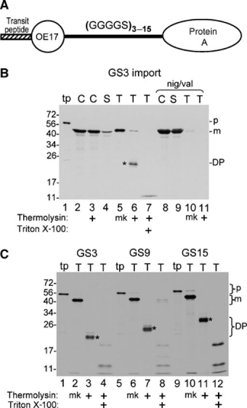
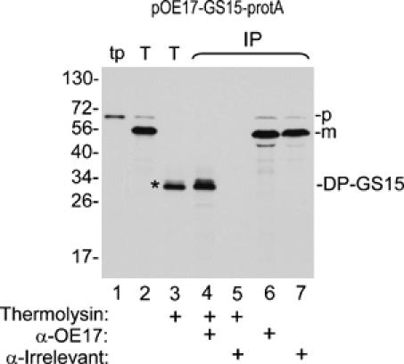
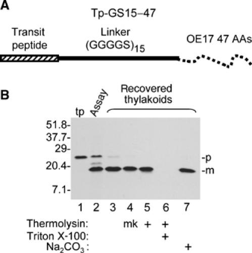
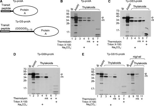
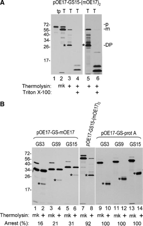
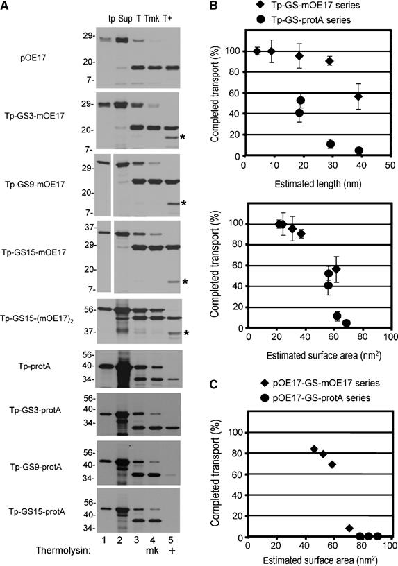
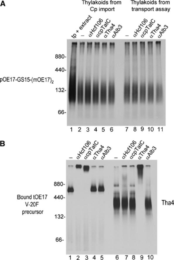
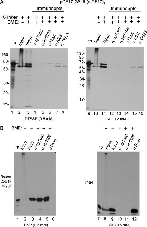
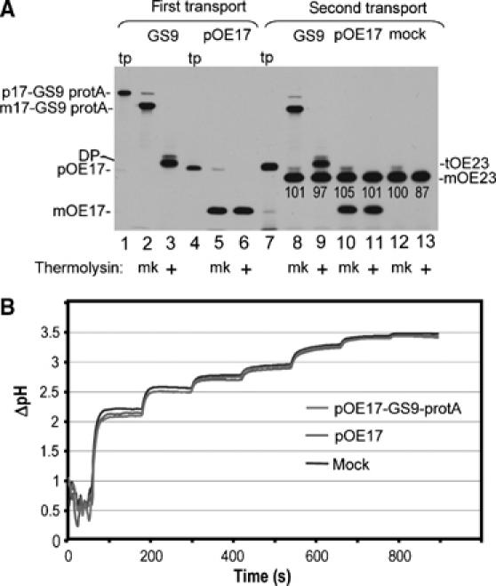
Tat systems transport completely folded proteins across ion-tight membranes. Three membrane proteins comprise the Tat machinery in most systems. In thylakoids, cpTatC and Hcf106 mediate precursor recognition, whereas Tha4 facilitates translocation. We used chimeric precursor proteins with unstructured peptides and folded domains to test predictions of competing translocation models. Two models invoke protein-conducting channels, whereas another model proposes that cpTatC pulls substrates through a patch of Tha4 on the lipid bilayer. The thylakoid system transported unstructured peptide substrates alone or when fused to folded domains. However, larger substrates stalled before completion, some with amino- and carboxyl-folded domains on opposite sides of the membrane. The length of the precursor that resulted in translocation arrest (20 to 30 nm) exceeded that expected for a single 'pull' mechanism, suggesting that a sustained driving force rather than a single pull moves the protein across the bilayer. Three different methods showed that stalled substrates were not stuck in a channel or even associated with Tat machinery. This finding favors the Tha4 patch model for translocation.


Figures

Figure 1
Figure 1
Chimeric precursor proteins containing pOE17, linkers consisting of repeats of the peptapeptide GGGGS, and protein A are arrested mid transport by the cpTat pathway. (A) Diagrammatic representation of the chimeric precursor proteins used in this experiment. (B) Import of pOE17-GS3-protA into intact chloroplasts. In vitro translated pOE17-GS3-protA (lane 1) was incubated with intact chloroplasts in an import assay, in the absence (lanes 2–7) or presence (lanes 8–11) of nigericin and valinomycin (Materials and methods). Recovered chloroplasts were either treated with thermolysin (lane 3) or not (lane 2), and intact chloroplasts repurified. The untreated chloroplasts were lysed and fractionated into a stromal fraction (lanes 4 and 9) and a thylakoid fraction (lanes 5 and 10). Aliquots of the thylakoid fraction were treated with thermolysin in the absence (lanes 6 and 11) or presence of 1% Triton X-100 (lane 7). (C) Transport of pOE17-GS(3–15)-protA into washed thylakoids. In vitro translated precursors (lanes 1, 5 and 9) were incubated with washed thylakoids in a transport assay (Materials and methods). Recovered thylakoids were mock treated (lanes 2, 6 and 10) or treated with thermolysin, in the absence (lanes 3, 7 and 11) or presence of 1% Triton X-100 (lanes 4, 8 and 12). Translation products (tp) represent 5% of assays; all other samples represent 100% of assays. Designations: p, precursor; m, mature form; DP, degradation product; mk, mock treatment. Asterisks indicate the protease resistant degradation products.
Figure 2
Figure 2
The OE17 moiety of pOE17-GS15-protA is transported across the thylakoid membrane, whereas the protein A moiety remains on the stromal side of the thylakoid membrane. Thylakoids recovered from a transport assay (Materials and methods) with pOE17-GS15-protA were thermolysin treated (lane 3) or not (lane 2), as shown below the panel, and were subjected to denaturing immunoprecipitation (IP, lanes 4–7) with antibodies to OE17, or an irrelevant antibody, as described in Materials and methods and depicted below the panel. Translation product (lane 1), 2% of assay; thylakoids and thermolysin-treated thylakoids, 50% of assays; eluates from the antibody beads, 100% of assays.
Figure 3
Figure 3
Thylakoid transport of the unstructured linker (GGGGS)15. (A) Diagrammatic representation of the precursor used for this experiment, which is called Tp-GS15-47 (see Materials and methods for details). (B) In vitro translated Tp-GS15-47 (lane 1) was assayed for thylakoid transport with chloroplast lysate for 30 min in the light. Lane 2 contains an aliquot of the assay mixture. Recovered thylakoids were analyzed directly (lane 3), mock treated (lane 4, mk), treated with thermolysin, in the absence (lane 5) or presence (lane 6) of 1% Triton X-100, or extracted with 0.2 M Na2CO3 (lane 7). Translation product (tp) represents 6% of assays, the aliquot of reaction mixture 40% of the assay, and other samples 100%.
Figure 4
Figure 4
Processing and transport of chimeric proteins consisting of the pOE17 transit peptide (Tp), linkers consisting of 3, 9, and 15 repeats of the GGGGS pentapeptide, and protein A. (A) Diagrammatic representation of the precursors used in this experiment. (B–E) Parallel washed thylakoid transport assays were conducted with Tp-protA (panel B), Tp-GS3-protA (panel C), Tp-GS9-protA (panel D), and Tp-GS15-protA (panel E), as described (Materials and methods). Aliquots of the assay mixtures were removed (lanes labeled assay) and the recovered thylakoids were analyzed directly, extracted with 0.2 M Na2CO3, mock treated (designated mk), or treated with thermolysin, in the absence or presence of 1% Triton X-100, as shown below the panels and described (Materials and methods). The assay in lanes 9–11 of panel E was conducted in the presence of nigericin and valinomycin. Translation product (tp) represents 7.5% of assays, the assay mixture, 50% of assays, and other samples, 100% of assays.
Figure 5
Figure 5
Transport of pOE17-GS-mOE17, pOE17-GS15-(mOE17)2, and pOE17-GS-protA into thylakoids. (A) pOE17-GS15-(mOE17)2 was incubated with washed thylakoids in a transport assay. Recovered thylakoids were either mock treated (mk) or treated with thermolysin, in the absence or presence of 1% Triton X-100, as shown below the panels. The gel in the right panel was exposed to film for three times as long as the left panel. (B) Concurrent assay of all precursors in parallel washed thylakoid transport assays. Recovered thylakoids were mock treated (mk) or treated with thermolysin, as shown. All panels were from the same gel, but exposed to film for different times, to adjust for different translation efficiencies. Numbers below the lanes represent the percentage of processed protein that was arrested mid transport. This was calculated from the DPM of the degradation product adjusted for the number of leucine residues in the mature protein versus degradation product. Translation products represent 6.5% and all other samples represent 100% of the assay mixtures.
Figure 6
Figure 6
Translocation completion as related to estimated substrate dimensions. (A) Example of transport assays used to calculate the percentage of completed translocation. Precursors were assayed for transport with washed thylakoids (Materials and methods), except that assays were 100 μl final volume and contained thylakoids equivalent to 50 μg chlorophyll. Thylakoids were recovered by centrifugation, the supernatant removed (Sup, lane 2), and the thylakoids analyzed directly (T, lane 3), mock treated (Tmk, lane 4), or treated with thermolysin (T+, lane 5). Translation products (tp, lane 1) represent 10% of the assay and all other samples 100%. (B) The percentage of completed transport was calculated from the amount of protease-protected processed substrate in lane 5 (e.g., m-GS-OE17 or m-GS-protA), compared to the processed substrate in lane 4 in panel A. This was plotted against the estimated length of the substrate (upper plot), or the estimated surface area (lower plot). The data are the means and standard error of the mean for three independent experiments, in which all substrates shown in panel A were assayed in parallel transport reactions (Supplementary Tables 1 and 2). The lengths used were as follows: OE17, 4.5 nm; protein A, 14 nm; and the GGGGS linkers, 0.33 nm per residue. Surface area was estimated as the surface of cylinders with the diameter of OE17 as 1.5 nm, protein A as 1.2 nm, and the GGGGS linker as 0.2 nm. (C) Plot of estimated surface area versus the percentage of completed transport of the pOE17-GS-X series in the experiment shown in Figure 5.
Figure 7
Figure 7
Attempt to detect association of arrested substrates with cpTat components by BN-PAGE and antibody mobility shift. Thylakoids recovered from a chloroplast import assay or thylakoid transport assay with pOE17-GS15-(mOE17)2, a binding assay with tOE17 V-20F, or an integration assay with Tha4 (Materials and methods), were solubilized with 0.75% digitonin and the soluble extracts (10 μl) incubated with 2 μl of buffer or 2 μl (8 μg) of IgG for 90 min on ice. Samples were then subjected to BN-PAGE and fluorography. (A) pOE17-GS15-(mOE17)2. Lane 1 contains 2 μl of translation product mixed with 10 μl of a digitonin extract of mock-incubated thylakoids. Lanes 2–6 contain solubilized membranes from the import assay incubated with buffer (lane 2) or IgGs, as shown above the panel. Lanes 7–11 contain solubilized membranes from the thylakoid transport assay incubated with buffer (lane 7) or IgGs, as shown. (B) Lanes 1–5 contain solubilized membranes from the tOE17 V-20F binding assay and lanes 6–10 contain solubilized membranes from the Tha4 integration assay. Molecular weight markers were ferritin (440 and 880 kDa) and bovine serum albumin (66 and 132 kDa). Protease treatment of thylakoids indicated that mOE17-GS15-(mOE17)2 was 77% transport-arrested in the import assay, and 81% arrested in the thylakoid transport assay.
Figure 8
Figure 8
Attempt to identify association of arrested substrates with cpTat components by chemical crosslinking and co-immunoprecipitation. Thylakoids from a transport assay with pOE17-GS15-(mOE17)2, a binding assay with tOE17 V-20F, or an integration assay with Tha4 were subjected to crosslinking with the indicated amounts of DTSSP or DSP (Materials and methods). Thylakoids were recovered, dissolved in 1% SDS, and subjected to immunoprecipitation (Materials and methods). Elution from the antibody beads was with SDS buffer containing β mercaptoethanol (BME) as shown above the panels to break the crosslinker. (A) Immunoprecipitation of crosslinked mOE17-GS15-(mOE17)2 was as designated above the panels. The relative amounts of samples loaded were as follows: translation product (tp, lanes 1 and 9), 4% of the transport assay; the input to the immunoprecipitations in the absence (lanes 2 and 10) or presence (lanes 3 and 11) of BME, 20% of the assay; and the antibody eluates, 100% of the assay. Thermolysin treatment of the thylakoids indicated that 90% of mOE17-GS15-(mOE17)2 was transport arrested. (B) Thylakoid-bound tOE17 V-20F and thylakoid integrated Tha4 were crosslinked with 0.5 mM DSP and subjected to immunoprecipitation with antibody beads as shown above the panels and described in (A). Sample loading was the same as in (A).
Figure 9
Figure 9
Transport-arrested pOE17-GS9-protA neither interferes with subsequent transport by the cpTat system, nor does it impair the ability to generate and maintain a thylakoidal ΔpH. (A) Chloroplast lysate was incubated with in vitro translated pOE17-GS9-protA (lane 1), pOE17 (lane 4), or mock TnT translation in the light. After 20 min, each assay was supplemented with additional precursor or mock translation extract, and the incubation continued for 15 min. Thylakoids were recovered by centrifugation and washed. Aliquots of thylakoids were analyzed directly (lanes 2 and 5) or thermolysin treated (lanes 3 and 6), or incubated with the cpTat precursor tOE23 (lane 7) in a second transport reaction for 15 min in the light (lanes 8–13). The amounts of pOE17-GS9-protA and pOE17 transported in the first transport incubation were determined by scintillation counting of extracted gel bands and quantitative immunoblotting to be ∼10 000 molecules of mOE17-GS9-protA per chloroplast equivalent and ∼17 000 molecules of mOE17 per chloroplast equivalent. The relative amounts of tOE23 transported in the second incubation (shown below the mOE23 band) were normalized to the amount present in the mock-treated thylakoid assay, which was set at 100. (B) Thylakoids from the first transport incubation (panel A) were examined for the ability to generate a ΔpH (Materials and methods). The light intensities in sequential steps were as follows: 1.5, 3, 5, 6.5, 14, 28, and 50 μE/m2-sec, respectively. Traces represent the average of two independent measurements.

References

    1. Alami M, Luke I, Deitermann S, Eisner G, Koch HG, Brunner J, Muller M (2003) Differential interactions between a twin-arginine signal peptide and its translocase in Escherichia coli. Mol Cell 12: 937–946 - PubMed
    1. Alder NN, Theg SM (2003) Energetics of protein transport across biological membranes. A study of the thylakoid DeltapH-dependent/cpTat pathway. Cell 112: 231–242 - PubMed
    1. Arnon DI (1949) Copper enzymes in isolated chloroplasts. Polyphenol oxidase in Beta vulgaris. Plant Physiol 24: 1–15 - PMC - PubMed
    1. Asai T, Shinoda Y, Nohara T, Yoshihisa T, Endo T (1999) Sec-dependent pathway and DeltapH-dependent pathway do not share a common translocation pore in thylakoidal protein transport. J Biol Chem 274: 20075–20078 - PubMed
    1. Balsera M, Arellano JB, Revuelta JL, de las Rivas J, Hermoso JA (2005) The 1.49 Å resolution crystal structure of PsbQ from photosystem II of Spinacia oleracea reveals a PPII structure in the N-terminal region. J Mol Biol 350: 1051–1060 - PubMed

Publication types

MeSH terms