. 2007 Jul 11;26(13):3250-9.
doi: 10.1038/sj.emboj.7601744. Epub 2007 Jun 14.

Tumour prevention by a single antibody domain targeting the interaction of signal transduction proteins with RAS

Affiliations

Tumour prevention by a single antibody domain targeting the interaction of signal transduction proteins with RAS

Tomoyuki Tanaka et al. EMBO J. .

Abstract

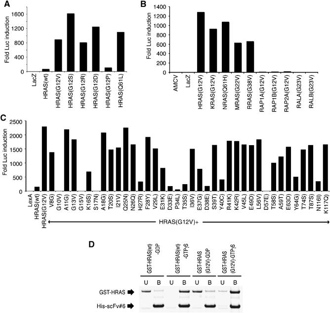
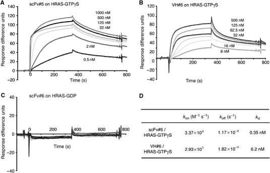
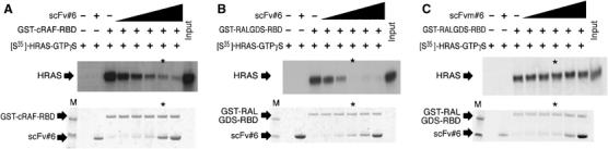
Many disease-related processes occur via protein complexes that are considered undruggable with small molecules. An example is RAS, which is frequently mutated in cancer and contributes to initiation and maintenance of the disease by constitutive signal transduction through protein interaction with effector proteins, like PI3K, RAF and RALGDS. Such protein interactions are therefore significant targets for therapy. We describe a single immunoglobulin variable region domain that specifically binds to activated GTP-bound RAS and prevents RAS-dependent tumorigenesis in a mouse model. The crystal structure of the immunoglobulin-RAS complex shows that the variable region competitively binds to the conformationally variant regions of RAS, where its signalling effector molecules interact. This allows the plasma membrane targeted single domain intrabody to inhibit signalling by mutant RAS. This mode of action is a novel advance to directly interfere with oncogenic RAS function in human cancer and shows a universally applicable approach to develop macromolecules to combat cancer. In addition, this method illustrates a general means for interfering with protein interactions that are commonly considered intractable as conventional drug targets.


Figures

Figure 1
Figure 1
Binding of the anti-RAS single domain with RAS proteins. The binding of the single VH domain to RAS was characterised using luciferase assays in COS-7 cells (A–C) or with bacterially expressed proteins (D). (A–C) The luciferase assays were conducted with a vector expressing iDab#6-VP16 fusion and various mutant forms of RAS, RAP1 or RAL as baits (The sequence alignments of RAS, RAP and RAL are shown in Supplementary Figure 2). (A) Assays with HRAS mutants, (B) RAS family mutants and (C) various mutants of the HRAS(G12V) backbone. (D) Interaction of RAS-GTPγS or -GDP and anti-RAS intrabody in vitro. Purified GST-RAS (wt) or GST-RAS(G12V) were loaded as GDP or GTPγS, as described in Materials and methods, and diluted into an Ni–agarose resin carrying purified His-tagged scFv#6. Following binding, the unbound (U) and bound (B) fractions were sampled and fractioned by SDS–PAGE.
Figure 2
Figure 2
The anti-RAS single domain reverts the RAS-transformed phenotype of mouse and human tumour cells. Retroviruses encoding iDab#6 or mutant (iDabm#6) iDab were infected into RAS-transformed mouse NIH3T3-EJ or DLD-1 and HT-1080 human cells. (A) A schematic diagram of retroviral constructs. To express anti-RAS single domain in mammalian cells, the cells were infected with ecotropic retrovirus (Costa et al, 2000). To localise the intrabody in the cells, a signal peptide was fused to the C-terminal of the anti-RAS intrabodies (either a nuclear localisation signal, nls or a plasma membrane targeting element, memb.). LTR, long terminal repeat; FLAG, antibody tag; IRES, internal ribosome entry site; SP, signal peptide (the sequences shown are for the nls or memb.). (B) The morphology of uninfected NIH3T3-D4 and NIH3T3-EJ are shown in top left and right panels and of NIH3T3-EJ cells infected with retrovirus encoding iDab#6-memb or mutant iDab#6 (iDabm#6-memb) in bottom left and right panels; images were obtained 48 h after infection. (C, D) Anchorage-independent growth of DLD-1 (C) and HT-1080 (D). The cells were infected with retrovirus expressing iDab#6 or retrovirus only. Forty-eight hours after infection, the EGFP expressing cells were sorted, seeded in soft agar and colonies (>0.1 mm) were counted at 3 weeks. Colony forming efficiency was the number of colonies per seeded cells. (E) Phalloidin staining of HT-1080 cells. Uninfected cells or cells infected with retrovirus encoding iDab#6 or iDabm#6 (left, middle or right) were stained 48 h after infection to show F-actin stress fibre formation.
Figure 3
Figure 3
The anti-RAS single domain inhibits tumorigenesis and metastasis in a mouse model. The effect of the anti-RAS single domain iDab#6 on the growth of mouse (NIH3T3-EJ; A, D) or human (HT-1080; B, or DLD-1; C) tumour cells was examined using grafts in immunodeficient nude mice. (A–C) Cells were infected with retroviral vectors expressing iDab#6 or the mutant iDabm#6, each with a membrane (memb.) localisation signal. Forty-eight hours after infection, the EGFP expressing cells were flow sorted, propagated in culture and injected subcutaneously into nude mice (5 × 10⁴ cells for NIH3T3-EJ, 2.5 × 10⁶ for HT-1080 or 5 × 10⁵ cells for DLD-1). If tumour sizes reached 17 mm diameter, the experiment was terminated, otherwise growth was observed for 42 days (for NIH3T3 cells) or 13 weeks (for HT-1080 or DLD-1). (D) Cells (105) were injected intravenously into nude mice. Three weeks after injection, the mice were killed, and lungs were dissected for examination. Representative images are shown from mice injected with either NIH3T3-D4 cells (top left), NIH3T3-EJ (top right), NIH3T3-EJ infected with retrovirus expressing iDab#6-memb (bottom left) or mutant iDabm#6-memb (bottom right).
Figure 4
Figure 4
Crystal structure of the RAS–anti-RAS single domains complex. HRAS(G12V) protein complexed with the anti-RAS #6 in an Fv format is shown in ribbon form (A) or space filling (B), where HRAS(G12V) is shown in green and the Fv proteins VH and VL are shown in cyan and orange, respectively. The CDRs of VH and VL are in yellow and lemon and the RAS switch I and II regions are in red and purple, respectively. The GTP and Mg2+ ion in RAS are in blue and magenta, respectively.
Figure 5
Figure 5
The binding site of the single domains on the RAS molecule. (A) A stereo diagram of the HRAS(G12V)-GTP-Fv binding interface. HRAS(G12V) is in green and the VH and VL chains are in cyan and orange, respectively. The CDRs of VH and VL are in yellow and lemon and the RAS switch I and II regions are in red and purple, respectively. Residues involved in the interface are shown in cylinder configuration. Specific residues of RAS are shown in blue, VH in red and VL in brown. Putative hydrogen bonds are indicated by dashed lines. (B) Schematic representation of the interacting residues in HRAS (green) and in the anti-RAS antibody (VH, yellow; VL, lemon). Putative hydrogen bonds are indicated by dotted lines. (C) The structures of HRAS(G12V)-GTP (green, red and purple) bound to anti-RAS Fv and of HRAS-GDP (blue) (PDB, 4Q21) (Milburn et al, 1990) are superimposed to illustrate the selectivity of iDab#6 single VH domain binding to activated GTP-bound RAS.
Figure 6
Figure 6
Affinity of anti-RAS intrabody for RAS. The binding affinity of the anti-RAS intrabody was measured using purified protein by surface resonance plasmon method measured using a BIAcore 2000. (A–C) Representative sensogrammes of scFv#6 bound to GST-HRAS(G12V)-GTPγS, bound to GST-HRAS(wt)-GDP (C), or of VH#6 bound to GST-RAS(G12V)-GTPγS are shown. The response difference units of sensogrammes, were normalised to the response of the channel trapping GST protein. (D) The table summarises values for the association (kon) (M−1 s−1) or dissociation rates (koff) (s−1), and the calculated equilibrium dissociation constants (Kd), using the BIAevaluation 2.1 software.
Figure 7
Figure 7
The anti-RAS single domain competes for binding of RAS effector molecules. (A–C) Competition of RAS-effector protein interaction by the anti-RAS intrabody in vitro. Biochemical studies of RAS-RAF or RAS-RALGDS interaction were performed using GST-fusion protein pull downs the presence of increasing amounts of anti-RAS scFv#6 (indicated by black shaded area)+presence, −absence. [35S-]HRAS(G12V) was loaded with GTPγS and mixed with differing concentrations of purified scFv#6 (A, B), mutant scFv#6 (C) plus by glutathione–Sepharose carrying GST-cRAF-RBD (A) or GST-RALGDS-RBD (B, C). The pulled down samples were eluted and fractionated by SDS–PAGE, followed by exposure to X-ray film, to detect [35S]-HRAS(G12V)-GTPγS (middle panel). The same amount of scFv and GST-RBD mixture without [35S]-HRAS(G12V)-GTPγS was fractioned on SDS–PAGE staining with CBB (lower panel). Asterisks indicate the lane in which scFv and GST-RBD were mixed in a 1:1 ratio. First lane (M) in lower panel is loaded with prestained SDS–PAGE standard molecular weight markers (estimated size: high, 53 kDa and low, 35.5 kDa).

References

    1. Adjei AA (2001) Blocking oncogenic Ras signaling for cancer therapy. J Natl Cancer Inst 93: 1062–1074 - PubMed
    1. Blundell TL, Sibanda BL, Montalvao RW, Brewerton S, Chelliah V, Worth CL, Harmer NJ, Davies O, Burke D (2006) Structural biology and bioinformatics in drug design: opportunities and challenges for target identification and lead discovery. Philos Trans R Soc Lond B Biol Sci 361: 413–423 - PMC - PubMed
    1. Canevari S, Biocca S, Figini M (2002) Re: blocking oncogenic Ras signaling for cancer therapy. J Natl Cancer Inst 94: 1031–1032, author reply 1032 - PubMed
    1. Cardinale A, Filesi I, Mattei S, Biocca S (2003) Evidence for proteasome dysfunction in cytotoxicity mediated by anti-Ras intracellular antibodies. Eur J Biochem 270: 3389–3397 - PubMed
    1. Cattaneo A, Biocca S (1997) Intracellular Antibodies: Development and Applications. Springer: New York, USA

Publication types

MeSH terms