

Valvulogenesis: the moving target
- PMID: 17569640
- PMCID: PMC2440410
- DOI: 10.1098/rstb.2007.2130
Valvulogenesis: the moving target
Abstract
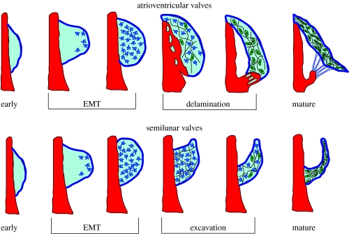
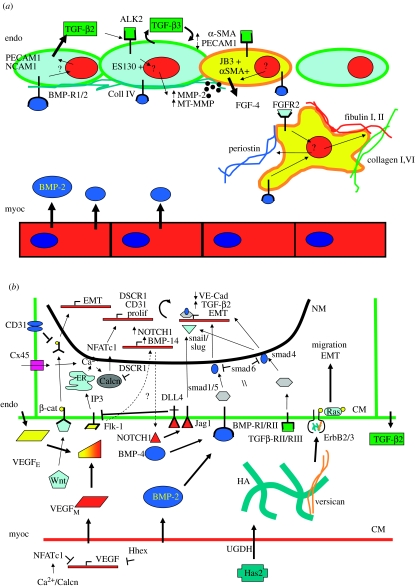
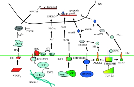
Valvulogenesis is an extremely complex process by which a fragile gelatinous matrix is populated and remodelled during embryonic development into thin fibrous leaflets capable of maintaining unidirectional flow over a lifetime. This process occurs during exposure to constantly changing haemodynamic forces, with a success rate of approximately 99%. Defective valvulogenesis results in impaired cardiac function and lifelong complications. This review integrates what is known about the roles of genetics and mechanics in the development of valves and how changes in either result in impaired morphogenesis. It is hoped that appropriate developmental cues and phenotypic endpoints could help engineers and clinicians in their efforts to regenerate living valve alternatives.
Figures

References
-
- Abdelwahid E, Rice D, Pelliniemi L.J, Jokinen E. Overlapping and differential localization of BMP-2, BMP-4, MSX-2 and apoptosis in the endocardial cushion and adjacent tissues of the developing mouse heart. Cell Tissue Res. 2001;305:67–78. doi:10.1007/s004410100399 - DOI - PubMed
-
- Adamo R.F, Guay C.L, Edwards A.V, Wessels A, Burch J.B. GATA-6 gene enhancer contains nested regulatory modules for primary myocardium and the embedded nascent atrioventricular conduction system. Anat. Rec. A Discov. Mol. Cell Evol. Biol. 2004;280:1062–1071. doi:10.1002/ar.a.20105 - DOI - PubMed
-
- Araki T, et al. Mouse model of Noonan syndrome reveals cell type- and gene dosage-dependent effects of Ptpn11 mutation. Nat. Med. 2004;10:849–857. doi:10.1038/nm1084 - DOI - PubMed
-
- Bajolle F, Zaffran S, Kelly R.G, Hadchouel J, Bonnet D, Brown N.A, Buckingham M. Rotation of the myocardial wall of the outflow tract is implicated in the normal positioning of the great arteries. Circ. Res. 2006;98:421–428. doi:10.1161/01.RES.0000202800.85341.6e - DOI - PubMed
-
- Baldwin H.S, Lloyd T.R, Solursh M. Hyaluronate degradation affects ventricular function of the early postlooped embryonic rat heart in situ. Circ. Res. 1994;74:244–252. - PubMed
Publication types
MeSH terms
LinkOut - more resources
Full Text Sources
Other Literature Sources