

Valvular endothelial cells and the mechanoregulation of valvular pathology
- PMID: 17569641
- PMCID: PMC2440407
- DOI: 10.1098/rstb.2007.2127
Valvular endothelial cells and the mechanoregulation of valvular pathology
Abstract
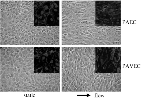
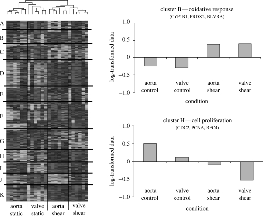
Endothelial cells are critical mediators of haemodynamic forces and as such are important foci for initiation of vascular pathology. Valvular leaflets are also lined with endothelial cells, though a similar role in mechanosensing has not been demonstrated. Recent evidence has shown that valvular endothelial cells respond morphologically to shear stress, and several studies have implicated valvular endothelial dysfunction in the pathogenesis of disease. This review seeks to combine what is known about vascular and valvular haemodynamics, endothelial response to mechanical stimuli and the pathogenesis of valvular diseases to form a hypothesis as to how mechanical stimuli can initiate valvular endothelial dysfunction and disease progression. From this analysis, it appears that inflow surface-related bacterial/thrombotic vegetative endocarditis is a high shear-driven endothelial denudation phenomenon, while the outflow surface with its related calcific/atherosclerotic degeneration is a low/oscillatory shear-driven endothelial activation phenomenon. Further understanding of these mechanisms may help lead to earlier diagnostic tools and therapeutic strategies.
Figures


References
-
- Affonso da Costa F.D, et al. The Ross procedure: is it the ideal operation for the young with aortic valve disease? Heart Surg. Forum. 1998;1:116–124. - PubMed
-
- Akinkugbe O.O, Nicholson G.D, Cruickshank J.K. Heart disease in blacks of Africa and the Caribbean. Cardiovasc. Clin. 1991;21:377–391. - PubMed
-
- Allen B.L, Katz B, Hook M. Streptococcus anginosus adheres to vascular endothelium basement membrane and purified extracellular matrix proteins. Microb. Pathog. 2002;32:191–204. doi:10.1006/mpat.2002.0496 - DOI - PubMed
-
- Allison M.A, Wright C.M. Age and gender are the strongest clinical correlates of prevalent coronary calcification (R1) Int. J. Cardiol. 2005;98:325–330. doi:10.1016/j.ijcard.2004.03.015 - DOI - PubMed
-
- Aluri S, Chandran K.B. Numerical simulation of mechanical mitral heart valve closure. Ann. Biomed. Eng. 2001;29:665–676. doi:10.1114/1.1385810 - DOI - PubMed
MeSH terms
LinkOut - more resources
Full Text Sources
Medical
