. 2007 Aug 10;282(32):23631-8.
doi: 10.1074/jbc.M701676200. Epub 2007 Jun 14.

Protein kinase Cepsilon (PKCepsilon) and Src control PKCdelta activation loop phosphorylation in cardiomyocytes

Affiliations

Protein kinase Cepsilon (PKCepsilon) and Src control PKCdelta activation loop phosphorylation in cardiomyocytes

Vitalyi O Rybin et al. J Biol Chem. .

Abstract

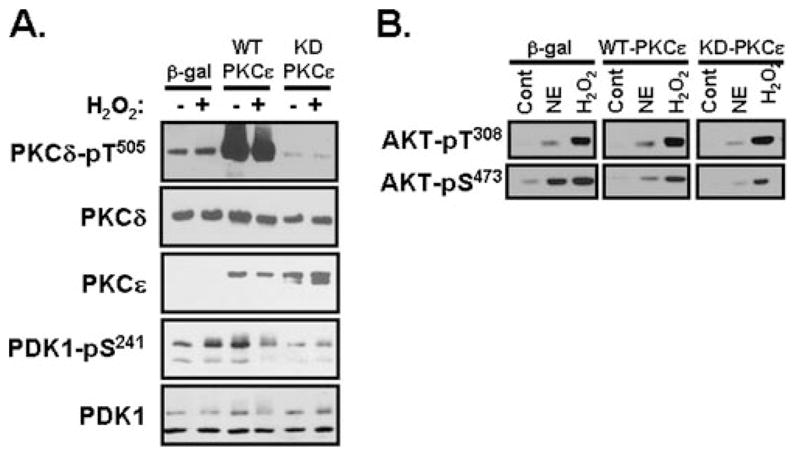
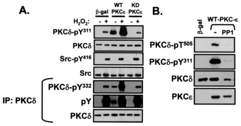
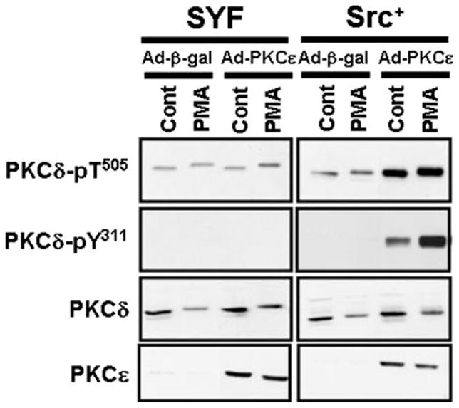
Protein kinase Cdelta (PKCdelta) is unusual among AGC kinases in that it does not require activation loop (Thr(505)) phosphorylation for catalytic competence. Nevertheless, Thr(505) phosphorylation has been implicated as a mechanism that influences PKCdelta activity. This study examines the controls of PKCdelta-Thr(505) phosphorylation in cardiomyocytes. We implicate phosphoinositide-dependent kinase-1 and PKCdelta autophosphorylation in the "priming" maturational PKCdelta-Thr(505) phosphorylation that accompanies de novo enzyme synthesis. In contrast, we show that PKCdelta-Thr(505) phosphorylation dynamically increases in cardiomyocytes treated with phorbol 12-myristate 13-acetate or the alpha(1)-adrenergic receptor agonist norepinephrine via a mechanism that requires novel PKC isoform activity and not phosphoinositide-dependent kinase-1. We used a PKCepsilon overexpression strategy as an initial approach to discriminate two possible novel PKC mechanisms, namely PKCdelta-Thr(505) autophosphorylation and PKCdelta-Thr(505) phosphorylation in trans by PKCepsilon. Our studies show that adenovirus-mediated PKCepsilon overexpression leads to an increase in PKCdelta-Thr(505) phosphorylation. However, this cannot be attributed to an effect of PKCepsilon to function as a direct PKCdelta-Thr(505) kinase, since the PKCepsilon-dependent increase in PKCdelta-Thr(505) phosphorylation is accompanied by (and dependent upon) increased PKCdelta phosphorylation at Tyr(311) and Tyr(332). Further studies implicate Src in this mechanism, showing that 1) PKCepsilon overexpression increases PKCdelta-Thr(505) phosphorylation in cardiomyocytes and Src(+) cells but not in SYF cells (that lack Src, Yes, and Fyn and exhibit a defect in PKCdelta-Tyr(311)/Tyr(332) phosphorylation), and 2) in vitro PKCdelta-Thr(505) autophosphorylation is augmented in assays performed with Src (which promotes PKCdelta-Tyr(311)/Tyr(332) phosphorylation). Collectively, these results identify a novel PKCdelta-Thr(505) autophosphorylation mechanism that is triggered by PKCepsilon overexpression and involves Src-dependent PKCdelta-Tyr(311)/Tyr(332) phosphorylation.


Figures

FIGURE 1
FIGURE 1. NE- and PMA-dependent PKCδ phosphorylation mechanisms in cardiomyocytes
Immunoblots of cell extracts from cardiomyocyte cultures pretreated for 45 min with vehicle, GF019023X (GFX; 5 μM in A and B or the indicated concentrations in C), Go6976 (5 μM) (A), LY294002 (LY; 10 μM) (A), or UCN-01 (0.1 μM in B, the indicated concentrations in C), followed by stimulations for 10 min with NE (10 μM), PMA (300 nM), or H2O2 (5 mM) as indicated. pT, phosphothreonine; pS, phosphoserine.
FIGURE 2
FIGURE 2. The controls of WT-PKCδ and KD-PKCδ Thr505 phosphorylation
Adenovirus-mediated gene transfer was used to overexpress WT-PKCδ and KD-PKCδ (each at MOI = 100) in the absence or presence of UCN-01 (0.3 μM). Cell extracts were then subjected to immunoblotting to compare PKCδ protein abundance and PKCδ-Thr505 (T505) phosphorylation. Since WT-PKCδ over-expression was 35 times higher than KD-PKCδ expression (at a similar MOI), protein loading was adjusted so that activation loop phosphorylation could be compared on similar amounts of heterologously overexpressed WT and KD enzymes. The figure shows that UCN-01 decreases WT-PKCδ-Thr505 phosphorylation and completely abrogates KD-PKCδ-Thr505 phosphorylation; KD-PKCδ-Thr(P)505 immunoreactivity was below the limits of detection in UCN-01-treated cultures, even when protein loading was increased and gel exposure times were prolonged
FIGURE 3
FIGURE 3. Ad-PKCε increases PKCδ-Thr505 phosphorylation without increasing PDK-1 expression, PDK-1 activity, or PKCδ protein abundance in cardiomyocytes
Adenovirus-mediated gene transfer was used to overex-press WT-PKCε, KD-PKCε, or β-galactosidase as a control in cardiomyocyte cultures (each at an MOI of 100 pfu/cell). 48 h following infection, cultures were treated for 10 min with vehicle, NE (10 μM) or H2O2 (5 mM) as indicated. Immunoblotting was with the indicated antibodies as described under “Experimental Procedures.” pT, phosphothreonine; pS, phosphoserine.
FIGURE 4
FIGURE 4. Ad-PKCε increases PKCδ-Thr505 (T505) phosphorylation without altering PKCδ (or PKCα) partitioning to membranes; Ad-PKCε slows PKCδ down-regulation
Adenovirus-mediated gene transfer was used to overexpress WT-PKCε or β-galactosidase as control (MOI of 100 pfu/cell). Cells were partitioned into soluble and particulate fraction 48 h postinfection following a 20-min challenge with vehicle or 300 nM PMA or a 24-h treatment with 300 nM PMA as indicated. Immunoblotting on soluble and particulate fractions was performed as described under “Experimental Procedures.”
FIGURE 5
FIGURE 5. Ad-PKCε increases PKCδ-Tyr311 and -Tyr332 phosphorylation without activating Src in cardiomyocytes
A, adenovirus-mediated gene transfer was used to overexpress WT-PKCε, KD-PKCε, or β-galactosidase as a control (MOI of 100 pfu/cell). 48 h following infection, cultures were treated for 10 min with vehicle or H2O2 (5 mM). Immunoblotting was with the indicated antibodies on cell extracts (top) or following PKCδ immunoprecipitation (IP) (bottom). B, WT-PKCε overexpression was in the absence or presence of PP1 (10 μM), with β-galactosidase as control. Immunoblotting was on cell extracts with the indicated antibodies as described under “Experimental Procedures.” pY, phosphotyrosine; T, threonine; pT, phosphothreonine.
FIGURE 6
FIGURE 6. Ad-PKCε increases PKCδ-Thr505 (T505) and -Tyr311 (Y311) phosphorylation in PKCε/& minus; MEFs and cardiac fibroblasts
Adenovirus-mediated gene transfer was used to overexpress WT-PKCε (at increasing MOI) in PKCε−/− MEFs and primary cardiac fibroblast cultures. Immunoblotting on lysates prepared 48 h following infection was with the indicated antibodies.
FIGURE 7
FIGURE 7. Src enhances in vitro PKCδ autophosphorylation at Thr505
In vitro kinase assays were performed with PKCδ and active Src as described under “Experimental Procedures.” Proteins were separated by SDS-PAGE and subjected to autoradiography and immunoblotting for PKCδ protein and phosphorylation. pT, phosphothreonine; pY, phosphotyrosine.
FIGURE 8
FIGURE 8. PKCε overexpression increases PKCδ-Tyr311 (pY311) and -Thr505 (pT505) phosphorylation in Src+ cells but not in SYF cells that lack Src, Yes, and Fyn expression
Shown is immunoblotting on cell extracts prepared from Src+ and SYF cells that were cultured and infected with Ad-PKCε or Ad-β-galactosidase and then treated with vehicle or PMA (300 nM for 20 min) as described under “Experimental Procedures.”

References

    1. Rybin VO, Sabri A, Short J, Braz JC, Molkentin JD, Steinberg SF. J Biol Chem. 2003;278:14555–14564. - PubMed
    1. Steinberg SF. Biochem J. 2004;384:449–459. - PMC - PubMed
    1. Le Good JA, Ziegler WH, Parekh DB, Alessi DR, Cohen P, Parker PJ. Science. 1998;281:2042–2045. - PubMed
    1. Stempka L, Schnolzer M, Radke S, Rincke G, Marks F, Gschwendt M. J Biol Chem. 1999;274:8886–8892. - PubMed
    1. Liu Y, Belkina NV, Graham C, Shaw S. J Biol Chem. 2006;281:12102–12111. - PubMed

Publication types

Substances