Evidence of intercellular coupling between co-cultured adult rabbit ventricular myocytes and myofibroblasts
- PMID: 17569734
- PMCID: PMC2277230
- DOI: 10.1113/jphysiol.2007.135038
Evidence of intercellular coupling between co-cultured adult rabbit ventricular myocytes and myofibroblasts
Abstract
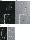
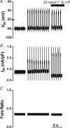
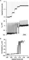
Intercellular coupling between ventricular myocytes and myofibroblasts was studied by co-culturing adult rabbit ventricular myocytes with previously prepared layers of cardiac myofibroblasts. Intercellular coupling was examined by: (i) tracking the movement of the fluorescent dye calcein; (ii) immunostaining for connexin 43 (Cx43); and (iii) measurement of intracellular [Ca2+] ([Ca2+]i). The effects of stimulating ventricular myocytes on the underlying myofibroblasts was examined by confocal measurements of [Ca2+]i using fluo-3. When ventricular myocytes were preloaded with calcein and co-cultured with myofibroblasts for 24 h, calcein fluorescence was detected in 52+/-4% (n=8 co-cultures) of surrounding myofibroblasts. Treatment with the gap junction uncoupler heptanol significantly reduced the movement of calcein (12+/-3%, n=6 co-cultures). Immunostaining showed expression of Cx43 in co-cultured myofibroblasts and myocytes. Field stimulation of ventricular myocytes co-cultured with myofibroblasts increased myofibroblast [Ca2+]i, no response was observed after treatment with heptanol or stimulation of fibroblasts in the absence of ventricular myocytes. Action potential parameters of ventricular myocytes in co-culture were similar to control values. However, application of the hormone sphingosine-1-phosphate (S-1-P) to the co-culture caused a depolarization of ventricular myocytes to approximately -20 mV. Sphingosine-1-phosphate had no effect on ventricular myocytes alone. Voltage-clamp measurements of isolated myofibroblasts indicated that S-1-P activated a significant quasi-linear current with a reversal potential of approximately -40 mV. In conclusion, this study shows that stimulation of the ventricular myocyte influences the intracellular Ca2+ of the linked myofibroblast via connexons. These intercellular links also allow the myofibroblasts to influence the electrical activity of the myocyte. This work indicates the nature of the gap junction-mediated bi-directional interactions that occur between ventricular myocyte and myofibroblast.
Figures


References
-
- Baudino T, Carver W, Giles WR, Borg TK. Cardiac fibroblasts: friend or foe. Am J Physiol Heart Circ Physiol. 2006;291:H1015–H1026. - PubMed
-
- Brilla CG, Scheer C, Rupp H. Angiotensin II and intracellular calcium of adult cardiac fibroblasts. J Mol Cell Cardiol. 1998;30:1237–1246. - PubMed
-
- Camelliti P, Devlin GP, Matthews KG, Kohl P, Green CR. Spatially and temporally distinct expression of fibroblast connexins after sheep ventricular infarction. Cardiovasc Res. 2004a;62:415–425. - PubMed
Publication types
MeSH terms
Substances
LinkOut - more resources
Full Text Sources
Other Literature Sources
Miscellaneous

