. 2007 Aug 15;583(Pt 1):315-27.
doi: 10.1113/jphysiol.2007.134726. Epub 2007 Jun 14.

Developmental changes in brain-derived neurotrophic factor-mediated modulations of synaptic activities in the pontine Kölliker-Fuse nucleus of the rat

Affiliations

Developmental changes in brain-derived neurotrophic factor-mediated modulations of synaptic activities in the pontine Kölliker-Fuse nucleus of the rat

Miriam Kron et al. J Physiol. .

Abstract

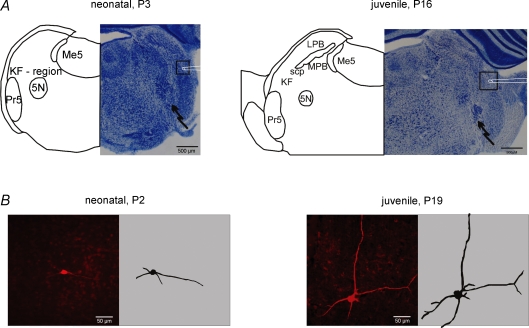
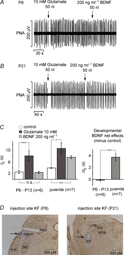
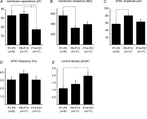
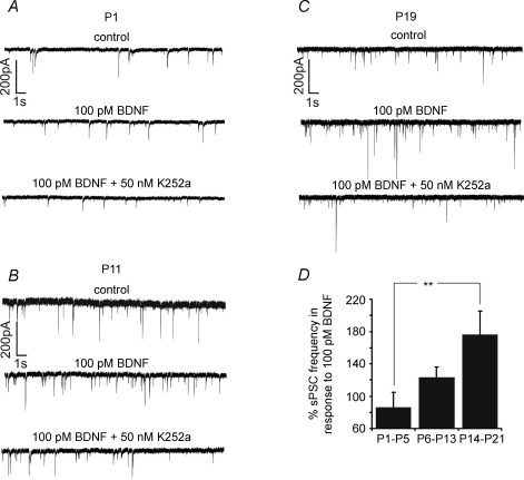
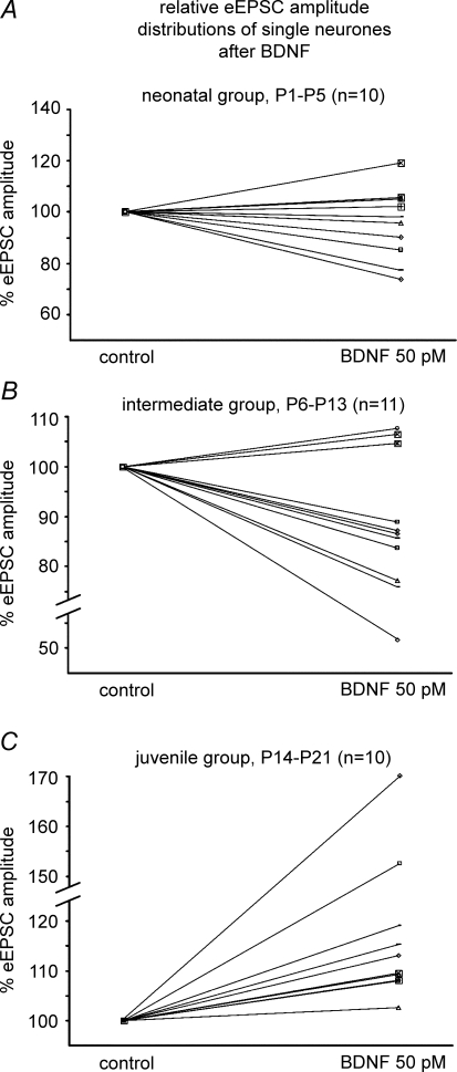
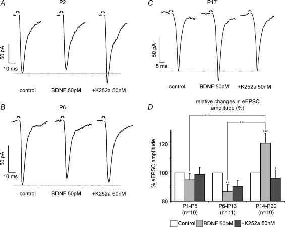
The Kölliker-Fuse nucleus (KF), part of the respiratory network, is involved in the modulation of respiratory phase durations in response to peripheral and central afferent inputs. The KF is immature at birth. Developmental changes in its physiological and anatomical properties have yet to be investigated. Since brain-derived neurotrophic factor (BDNF) is of major importance for the maturation of neuronal networks, we investigated its effects on developmental changes in the KF on different postnatal days (neonatal, P1-5; intermediate, P6-13; juvenile, P14-21) by analysing single neurones in the in vitro slice preparation and network activities in the perfused brainstem preparation in situ. The BDNF had only weak effects on the frequency of mixed excitatory and inhibitory spontaneous postsynaptic currents (sPSCs) in neonatal slice preparations. Postnatally, in the intermediate and juvenile age groups, a significant augmentation of the sPSC frequency was observed in the presence of 100 pm BDNF (+23.5+/-12.6 and +76.7+/-28.4%, respectively). Subsequent analyses of BDNF effects on evoked excitatory postsynaptic currents (eEPSCs) revealed significant enhancement of eEPSC amplitude of +20.8+/-7.0% only in juvenile stages (intermediates, -13.2+/-4.8%). On the network level, significant modulation of phrenic nerve activity following BDNF microinjection into the KF was also observed only in juveniles. The data suggest that KF neurones are subject to BDNF-mediated fast synaptic modulation after completion of postnatal maturation. After maturation, BDNF contributes to modulation of fast excitatory neurotransmission in respiratory-related KF neurones. This may be important for network plasticity associated with the processing of afferent information.


Figures

Figure 1
Figure 1
Developmental changes in cell mormphology of Kölliker–fuse neurones and cytoarchitecture of the dorsolateral pons A illustrates recording (square with schematic patch pipette) and stimulation sites (jagged arrow) in pontine slices of different postnatal stages. Please note the developing complexity of the cyto-architecture illustrated by thionin staining (right panels) and semischematic drawings (left panels). B shows photomicrographs of biocytin-stained KF neurones from the neonate and juvenile groups (left panels) and their two-dimensional reconstructions (right panels). Please note the increasing complexity of the dendritic tree during the postnatal maturation. Abbreviations: 5N, motor trigeminal nucleus; KF, Kölliker–Fuse nucleus; LPB, lateral parabrachial nucleus; Me5, mesencephalic trigeminal nucleus; MPB, medial parabrachial nucleus; Pr5, principal sensory trigeminal nucleus; and scp, superior cerebellar peduncle.
Figure 8
Figure 8
Illustration of respiratory modulation evoked by glutamate and BDNF microinjections into the KF in the perfused brainstem preparation derived from different postnatal stages A Illustrates that microinjections of glutamate in the intermediate group evoked apnoea while subsequent BDNF injection was ineffective. In contrast, at juvenile stages (B) BDNF evoked brief apnoeas similar to those observed after the preceding glutamate injections. C, bar graphs summarize the statistical analysis of the glutamate- and BDNF-evoked prolongation of the expiratory interval (tE) in the intermediate and juvenile age group (left). Developmental comparison of the BDNF-induced effect on tE is shown in the right diagram. *P < 0.05, ***P < 0.001. D, photomicrographs illustrating examples of the localization of injection sites for the intermediate and juvenile groups. Abbreviations: PNA, phrenic nerve activity; 5N, motor trigeminal nucleus; KF, Kölliker–Fuse nucleus; ll, lateral lemniscus; LPB, lateral parabrachial nucleus; Me5, mesencephalic trigeminal nucleus; MPB, medial parabrachial nucleus; Pr5, principal sensory trigeminal nucleus; scp, superior cerebellar peduncle.
Figure 2
Figure 2
Summary of developmental changes of membrane capacitance (A), membrane resistance (B), sPSC amplitude (C), sPSC frequency (D) and current density E) *P < 0.05, **P < 0.01.
Figure 3
Figure 3
Representative recordings of sPSC frequencies of a P1 (A), P11 (B) and P19 neurone (C) in response to BDNF (100 pm) and K252a (50 nm); the diagram (D) summarizes the developmental comparison of the effects of BDNF on sPSC frequency **P < 0.01.
Figure 4
Figure 4
Illustration of BDNF-mediated effects on sPSC frequency (left panels) and amplitude (right panels) after bath application of the drug at different postnatal stages The left panels of A, B and C summarize the dose–response curves of sPSC frequencies to different concentrations of BDNF. The right panels of A, B and C show that neither BDNF nor K252a had significant effects on the amplitude of sPSCs in the different developmental stages. *P < 0.05, ***P < 0.001.
Figure 5
Figure 5
Summary of BDNF-mediated effects on eEPSCs at different postnatal stages A, B and C illustrate the BDNF-induced modulation of eEPSC amplitudes from single neurones of the different developmental stages.
Figure 6
Figure 6
A, B and C show single examples of BDNF-mediated modulation of eEPSC amplitude at the neonate, the intermediate and the juvenile stage, repectively, while D illustrates developmental group data of BDNF and K252a effects on eEPSC amplitudes *P < 0.05, **P < 0.01, ***P < 0.001.
Figure 7
Figure 7
Developmental changes in weighted time constants and decay kinetics A illustrates BDNF-evoked effects on the weighted time constant (τw) in the different experimental groups. The BDNF had no significant effects on the decay of eEPSCs, but decay kinetics became significantly faster with increasing postnatal age. *P < 0.05. B shows an overlay of normalized currents before and after BDNF application to indicate no BDNF-evoked changes in decay kinetics.

Similar articles

Cited by

References

    1. Alheid GF, Milsom WK, McCrimmon DR. Pontine influences on breathing: an overview. Respir Physiol Neurobiol. 2004;143:105–114. - PubMed
    1. Aoki C, Wu K, Elste A, Len G, Lin S, McAuliffe G, Black IB. Localization of brain-derived neurotrophic factor and TrkB receptors to postsynaptic densities of adult rat cerebral cortex. J Neurosci Res. 2000;59:454–463. - PubMed
    1. Arvanian VL, Mendell LM. Acute modulation of synaptic transmission to motoneurons by BDNF in the neonatal rat spinal cord. Eur J Neurosci. 2001a;14:1800–1808. - PubMed
    1. Arvanian VL, Mendell LM. Removal of NMDA receptor Mg2+ block extends the action of neurotrophin-3 on synaptic transmission in neonatal rat motoneurons. J Neurophysiol. 2001b;86:123–129. - PubMed
    1. Baker-Herman TL, Fuller DD, Bavis RW, Zabka AG, Golder FJ, Doperalski NJ, Johnson RA, Watters JJ, Mitchell GS. BDNF is necessary and sufficient for spinal respiratory plasticity following intermittent hypoxia. Nat Neurosci. 2004;7:48–55. - PubMed

Publication types

Substances