. 2007 Aug;171(2):415-27.
doi: 10.2353/ajpath.2007.070075. Epub 2007 Jun 14.

Synaptopodin protects against proteinuria by disrupting Cdc42:IRSp53:Mena signaling complexes in kidney podocytes

Affiliations

Synaptopodin protects against proteinuria by disrupting Cdc42:IRSp53:Mena signaling complexes in kidney podocytes

Etsuko Yanagida-Asanuma et al. Am J Pathol. 2007 Aug.

Abstract

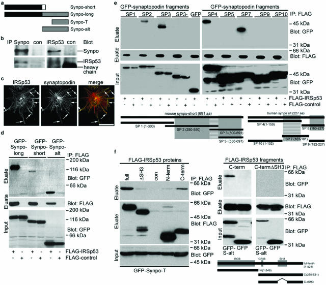
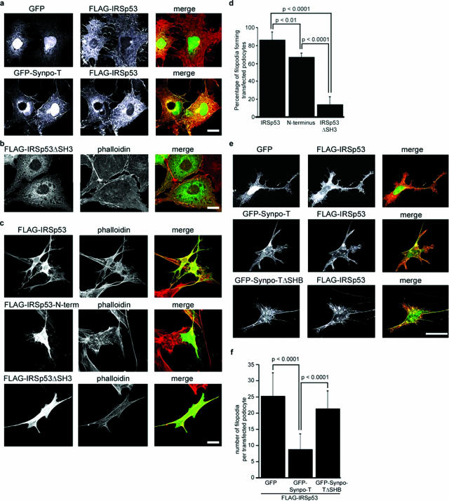
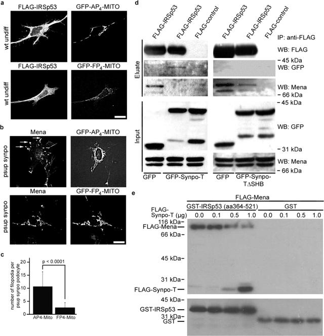
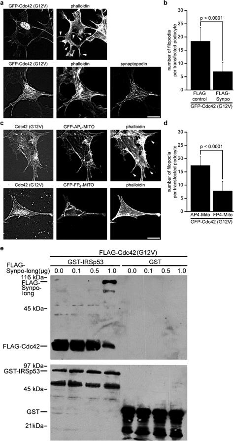
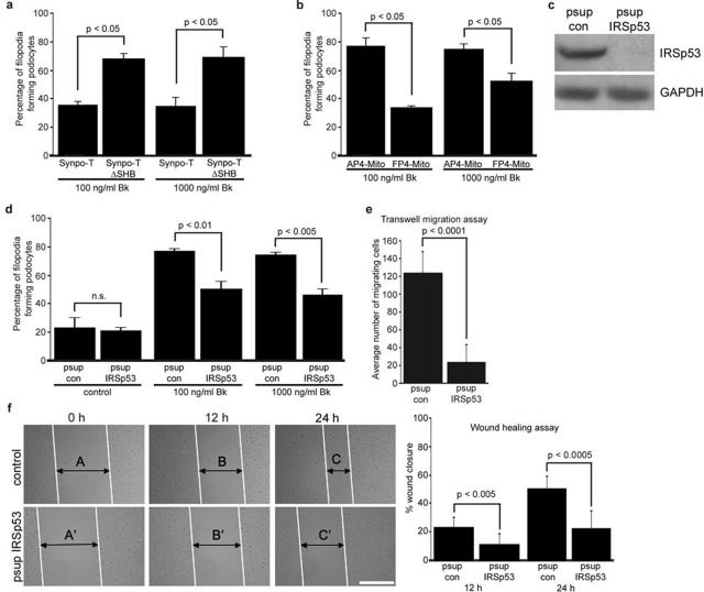
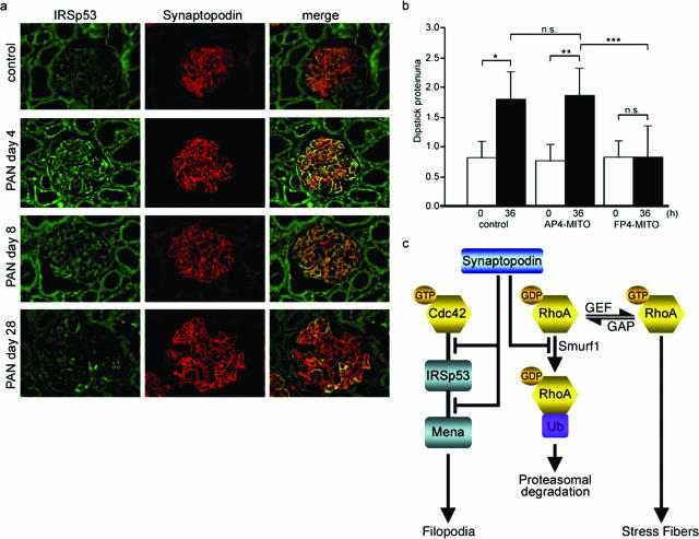
The actin-based foot processes of kidney podocytes and the interposed slit diaphragm form the final barrier to proteinuria. Mutations affecting several podocyte proteins cause disruption of the filtration barrier and rearrangement of the highly dynamic podocyte actin cytoskeleton. Proteins regulating the plasticity of the podocyte actin cytoskeleton are therefore of critical importance for sustained kidney barrier function. Synaptopodin is an actin-associated protein essential for the integrity of the podocyte actin cytoskeleton because synaptopodin-deficient mice display impaired recovery from protamine sulfate-induced foot process effacement and lipopolysaccharide-induced nephrotic syndrome. Moreover, bigenic heterozygosity for synaptopodin and CD2AP is sufficient to induce spontaneous proteinuria and focal segmental glomerulosclerosis-like glomerular damage in mice. Mechanistically, synaptopodin induces stress fibers by blocking the proteasomal degradation of RhoA. Here we show that synaptopodin directly binds to IRSp53 and suppresses Cdc42:IRSp53:Mena-initiated filopodia formation by blocking the binding of Cdc42 and Mena to IRSp53. The Mena inhibitor FP(4)-Mito suppresses aberrant filopodia formation in synaptopodin knockdown podocytes, and when delivered into mice protects against lipopolysaccharide-induced proteinuria. The identification of synaptopodin as an inhibitor of Cdc42:IRSp53:Mena signaling defines a novel antiproteinuric signaling pathway and offers new targets for the development of antiproteinuric therapeutic modalities.


Figures

Figure 1
Figure 1
Synaptopodin is a novel IRSp53-interacting protein. a: Schematic of synaptopodin isoforms. The Synpo-alt fragment of Synpo-long was used as bait construct in the two-hybrid screen and identified IRSp53 as synaptopodin-interacting protein. b: Coimmunoprecipitation experiments showing that endogenous synaptopodin interacts with endogenous IRSp53 in podocytes. Left: IP with anti-synaptopodin (Synpo). Right: IP with anti-IRSp53. No interaction is found with a control antibody against GFP (con). c: Deconvolution microscopy shows that IRSp53 and synaptopodin partially colocalize (arrows) in differentiated wild-type podocytes. d: GFP-tagged Synpo-long, Synpo-short, and Synpo-alt but not GFP alone coprecipitate with FLAG-IRSp53 from co-transfected HEK cells. e: Coimmunoprecipitation studies of FLAG-IRSp53 and GFP-tagged synaptopodin fragments SP1-SP10. Left: Synpo-short contains one interaction site for IRSp53 (striped box; amino acids 500 to 550) located between the α-actinin binding sites (gray boxes). SP1 and GFP alone do not interact with IRSp53. Right: Synpo-alt contains an independent IRSp53 binding site (striped box, amino acids 103 to 159), which overlaps with the first α-actinin binding site (gray boxes) of Synpo-alt. f: Left: Cotransfection of GFP-Synpo-T and various FLAG-tagged IRSp53 proteins. The strongest binding is seen with full-length IRSp53 (full) followed by the C-terminal fragment (C-term). A significantly weaker binding is found with IRSp53 lacking the SH3 domain (ΔSH3) or with an N-terminal fragment (N-term). No binding is seen with the FLAG control (con). Right: Cotransfection of GFP-Synpo-alt (S-alt) or GFP and FLAG-tagged IRSp53 fragments C-term or C-termΔSH3. Synaptopodin does not bind to C-termΔSH3. Altogether, IRSp53 contains two independent synaptopodin-binding sites. The first, weaker site overlaps with the RCB domain, the second, stronger site is the SH3 domain of IRSp53. Scale bar = 25 μm.
Figure 2
Figure 2
Synaptopodin inhibits IRSp53:Mena-induced filopodia. a: Top: Cotransfection of FLAG-IRSp53 and GFP induces filopodia in COS-7 cells. Bottom: GFP-Synpo-T (as well as Synpo-short, Synpo-long, or Synpo-alt; data not shown) suppresses IRSp53-induced filopodia. b: Overexpression of IRSp53ΔSH3 does not induce filopodia in transfected COS-7 cells. c: Induction of filopodia in transfected undifferentiated wild-type podocytes by FLAG-IRSp53 (top) and the truncated FLAG-N-terminal fragment (middle). Virtually no filopodia are seen with IRSp53ΔSH3 (bottom). d: Quantitative analysis of filopodia formation in transfected undifferentiated wild-type podocytes. See Results for details. e: Top: cotransfection of FLAG-IRSp53 and GFP induces filopodia in undifferentiated podocytes. Middle: GFP-Synpo-T suppresses IRSp53-induced filopodia. Bottom: GFP-Synpo-TΔSHB does not suppress IRSp53 filopodia. f: Quantitative analysis of synaptopodin-mediated inhibition of IRSp53-induced filopodia formation in podocytes. See Results for details. Scale bars = 25 μm.
Figure 3
Figure 3
Synaptopodin suppresses filopodia formation by disrupting IRSp53:Mena signaling complexes. a: Cotransfection with FP4-Mito but not AP4-Mito suppresses IRSp53-induced filopodia in undifferentiated wild-type (wt undiff) podocytes. b: In GFP-AP4-Mito-transfected, synaptopodin-knockdown (psup synpo) podocytes, Mena is targeted to aberrant filopodia (arrows). In contrast, GFP-FP4-Mito sequesters Mena to the mitochondrial surface and suppresses filopodia formation. c: Quantitative analysis of FP4-Mito-mediated inhibition of filopodia formation in synaptopodin knockdown podocytes. See Results for details. d: Left: Coimmunoprecipitation of endogenous Mena with FLAG-IRSp53 from COS-7 cells transfected with FLAG-IRSp53 and GFP. Cotransfection with GFP-Synpo-T blocks the binding of Mena to FLAG-IRSp53. Instead, GFP-Synpo-T coprecipitates with FLAG-IRSp53. No binding is found with a FLAG control. Right: Restored coimmunoprecipitation of endogenous Mena with FLAG-IRSp53 from COS-7 cells cotransfected with FLAG-IRSp53 and the GFP-Synpo-T mutant lacking the IRSp53 binding site (GFP-Synpo-TΔSHB). e: Immobilized GST-IRSp53 (amino acids 364 to 521) but not GST alone directly binds to purified FLAG-Mena. In the presence of increasing amounts of FLAG-Synpo-T, the binding of IRSp53 to Mena is gradually lost, whereas increased binding of Synpo-T to IRSp53 can be detected.
Figure 4
Figure 4
Synaptopodin blocks Cdc42:Mena-dependent filopodia formation by disrupting Cdc42:IRSp53 signaling complexes. a: Cotransfection with synaptopodin (bottom) suppresses the formation of Cdc42-induced filopodia (arrowheads) in undifferentiated wild-type podocytes (top). b: Quantitative analysis of synaptopodin-mediated inhibition of Cdc42-induced filopodia formation in undifferentiated wild-type podocytes. See Results for details. c: Cotransfection with FP4-Mito but not AP4-Mito suppresses Cdc42-induced filopodia (arrowheads) in undifferentiated wild-type podocytes. d: Quantitative analysis of FP4-Mito-mediated inhibition of Cdc42-induced filopodia formation in undifferentiated wild-type podocytes. See Results for details. e: Immobilized GST-IRSp53 (full-length) but not GST alone directly binds to purified FLAG-Cdc42. In the presence of increasing amounts of FLAG-Synpo-long, the binding of IRSp53 to Cdc42 is gradually reduced, whereas increasing binding of Synpo-long to IRSp53 can be observed. Scale bars = 25 μm.
Figure 5
Figure 5
Synaptopodin inhibits Bk-induced, IRSp53:Mena-mediated filopodia formation in podocytes. a: Synpo-T but not Synpo-TΔSHB, which cannot block the binding of Mena to IRSp53, reduces Bk-induced filopodia formation in transfected undifferentiated wild-type podocytes. See Results for details. b: FP4-Mito, but not the AP4-Mito control, reduces Bk-induced filopodia formation in undifferentiated wild-type podocytes. See Results for details. c: Western blot analysis of IRSp53 after gene silencing by stable expression of an IRSp53-specific siRNA (psup IRSp53). psup IRSp53 but not a control siRNA construct (psup con) suppresses IRSp53 protein expression in podocytes. d: Knockdown of IRSp53 significantly impairs Bk-induced filopodia formation in podocytes. e: Compared with wild-type podocytes, IRSp53 knockdown podocytes display significantly impaired motility in Transwell migration experiments. f: Scrape wound assay. In the absence of IRSp53, wound closure is significantly delayed at 12 and 24 hours. Compared with wild-type cells, IRSp53 knockdown podocytes display significantly impaired directed cell migration. See Results for details. Scale bar = 200 μm.
Figure 6
Figure 6
FP4-Mito ameliorates proteinuria by blocking IRSp53:Mena signaling. a: Transient up-regulation of IRSp53 protein expression in podocytes during PAN nephrosis. In PBS controls, only weak glomerular IRSp53 labeling is observed. On day 4 after PAN injection, IRSp53 expression is significantly increased in podocytes as shown by co-localization with synaptopodin. IRSp53 immunostaining in podocytes starts to decline on day 8 and returns to control levels on day 28. b: In vivo gene transfer of FP4-Mito but not AP4-Mito or luciferase (control) protects against LPS-induced proteinuria. *P = 0.000047, **P = 0.000014, ***P = 0.00039; n.s., not significant. c: A model for the antagonistic regulation of Cdc42 and RhoA signaling by synaptopodin. Synaptopodin suppresses IRSp53:Mena-mediated filopodia by blocking the binding of Cdc42 and Mena to IRSp53. In addition, synaptopodin induces stress fibers by competitive blocking of Smurf-1-mediated ubiquitination of RhoA, thereby preventing the targeting of RhoA for proteasomal degradation.

Similar articles

Cited by

References

    1. Mundel P, Kriz W. Structure and function of podocytes: an update. Anat Embryol (Berl) 1995;192:385–397. - PubMed
    1. Somlo S, Mundel P. Getting a foothold in nephrotic syndrome. Nat Genet. 2000;24:333–335. - PubMed
    1. Drenckhahn D, Franke RP. Ultrastructural organization of contractile and cytoskeletal proteins in glomerular podocytes of chicken, rat, and man. Lab Invest. 1988;59:673–682. - PubMed
    1. Kerjaschki D. Caught flat-footed: podocyte damage and the molecular bases of focal glomerulosclerosis. J Clin Invest. 2001;108:1583–1587. - PMC - PubMed
    1. Oh J, Reiser J, Mundel P. Dynamic (re)organization of the podocyte actin cytoskeleton in the nephrotic syndrome. Pediatr Nephrol. 2004;19:130–137. - PubMed

Publication types

MeSH terms